coalescent mastoiditis
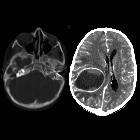
School ager
with pus draining from the right ear. Axial CT with contrast of the brain with bone windows (left) shows opacification and destruction of the right mastoid air cells while axial CT with contrast of the brain with soft tissue windows (right) shows a large low density ring enhancing lesion in the right cerebral hemisphere that is causing midline shift to the left.The diagnosis was right coalescent mastoiditis with an intracranial abscess.
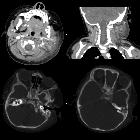
Infant with
left otitis media and left neck swelling. Axial and coronal CT with contrast of the neck with soft tissue windows (above) show extensive left cervical adenopathy and inflammation. Axial CT with contrast of the neck with bone windows (below) show bilateral complete opacification of the mastoid air cells and subtle erosive changes in the anterior aspect of the left temporal bone. There were no intracranial findings.The diagnosis was left coalescent mastoiditis.

Coalescent
mastoiditis • Mastoiditis - Ganzer Fall bei Radiopaedia

Cerebral
venous thrombosis, subdural empyema and cerebral abscess as complications of coalescent otomastoiditis. Coronal CT bone window showing right-sided mastoid air cell and middle ear occupation.

Cerebral
venous thrombosis, subdural empyema and cerebral abscess as complications of coalescent otomastoiditis. Axial CT bone window showing right-sided mastoid air cell and middle ear occupation. Erosion of mastoid air cells, resulting in communication with middle cerebral fossa.

Cerebral
venous thrombosis, subdural empyema and cerebral abscess as complications of coalescent otomastoiditis. Annotated image indicating the presence of occupation of mastoid air cell (orange arrow) and mastoid cortex erosion (blue arrow).

External and
middle ear diseases: radiological diagnosis based on clinical signs and symptoms. Acute coalescent otomastoiditis (ACOM). Axial scan in bone window demonstrates diffuse opacification of middle ear and mastoid with destructions of mastoid septations and sigmoid plate (arrow)
Coalescent mastoiditis is simply the term given to acute otomastoiditis when mucoperiosteal disease extends to involve the bone. The septae which normally separate one mastoid air cell from another are resorbed. This change is only easily appreciated on thin section bone-algorithm through the temporal bones. Comparison to the contralateral mastoid may be useful in detecting early changes, although normal asymmetry should be taken into account.
Complications (see acute otomastoiditis article) are much more likely once infection spreads to the bone, and include periauricular cellulitis and intracranial extension.
Siehe auch:
und weiter:


 Assoziationen und Differentialdiagnosen zu einschmelzende Mastoiditis:
