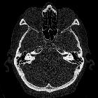
cochlear anomalies (classification)
Inner ear malformations are a spectrum of congenital anomalies involving the inner ear structures with an emphasis on the cochlea due to their implications for sensorineural hearing loss.
Classification
An imaging-based classification was first proposed in 1987 by Jackler et al. according to polytomography findings and embryological concepts . Using CT, Sennaroglu et al. later refined the classification and its pathophysiological basis . The latest (2017) iteration of Sennaroglu's classification, which is the most used worldwide, is as follows :
Items 1 through 6 above represent a spectrum ordered from the least to most differentiated inner ear structures due to the time of developmental arrest ranging between the 3week (for complete labyrinthine aplasia) and 7 week (for incomplete partition type II), with the exception that incomplete partition type I is posited to occur earlier than cochlear hypoplasia and common cavity is sometimes listed earlier than cochlear aplasia .
The above classification does not separately account for findings that may occur in association with other inner ear malformations but are sometimes isolated :
- vestibule dilation
- semicircular canal dysplasia
- internal auditory canal abnormalities
In addition to the above categories of findings, which are primarily based on CT, abnormalities of the cochlear nerve are important to classify by MRI :
- hypoplastic or absent cochlear nerve
- hypoplastic or absent common cochleovestibular nerve
Radiology report
Because of the complex and variable associations of individual components of these inner ear malformations, some have proposed a finely descriptive approach to reporting. In the INCAV classification system , imaging abnormalities are graded by severity for each of the major inner ear structures: internal auditory canal (I), cochlear nerve on MRI or cochlear nerve canal on CT (N), cochlea (C), vestibular aqueduct (A), and vestibule (V). For example, the cochlea may be classified as normal (0), incomplete partition type II (1), incomplete partition type III (2), hypoplasia (3), incomplete partition type I (4), common cavity (5), or aplasia (6).
Siehe auch:

 Assoziationen und Differentialdiagnosen zu Congenital inner ear anomalies:

