Colles fracture


Colles fractures are very common extra-articular fractures of the distal radius that occur as the result of a fall onto an outstretched hand. They consist of a fracture of the distal radial metaphyseal region with dorsal angulation and impaction, but without the involvement of the articular surface. This article describes radiographic features to check for and possible complications.
Epidemiology
Colles fractures are the most common type of distal radial fracture and are seen in all adult age groups and demographics. They are particularly common in patients with osteoporosis, and as such, they are most frequently seen in elderly women. The relationship between Colles fractures and osteoporosis is strong enough that when an older male patient presents with a Colles fracture, he should be investigated for osteoporosis because his risk of a hip fracture is also elevated .
Younger patients who sustain Colles fractures have usually been involved in high impact trauma or have fallen, e.g. during contact sports, skiing, horse riding .
Mechanism
Most Colles fractures are secondary to a fall on an outstretched hand (FOOSH) with a pronated forearm in dorsiflexion (the position one adopts when trying to break a forward fall).
The proximal row of the carpus (particularly the lunate and scaphoid) transfer energy to the distal radius, both in the dorsal direction and along the long axis of the radius. Most fractures are therefore dorsally angulated and impacted.
Radiographic features
A number of classification systems exist for distal forearm fractures. One of the more popular is the Frykman classification system, although it fails to distinguish between Smith and Colles fractures as it is based on AP radiographs . As such, in clinical practice, the use of the term Colles fracture with an appropriate description of any associated injuries is sufficient in most instances.
Plain films usually suffice, although if there is a concern of intra-articular extension, then CT may be beneficial.
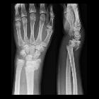
Plain radiograph
The plain radiographic series often comprises an AP and a lateral view; however, it is not uncommon for an oblique view to be included. The fracture appears extra-articular and usually proximal to the radioulnar joint. Dorsal angulation of the distal fracture fragment is present to a variable degree (as opposed to volar angulation of a Smith fracture). If dorsal angulation is severe enough, a dinner fork deformity may be described. There is also usually impaction with resultant shortening of the radius. An associated ulnar styloid fracture is present in up to 50% of cases.
Report checklist
In addition to noting the presence of a fracture a number of features should be sought and commented upon:
- fracture
- the degree of dorsal angulation
- the degree of impaction
- degree and direction of displacement
- location of the medial fracture line: does it involve the radioulnar joint
- presence of intra-articular fractures
- other fractures
- ulnar styloid
- carpal bones
Treatment and prognosis
The vast majority of Colles fractures can be treated with closed reduction and cast immobilization. The cast extends from below the elbow to the metacarpal heads and holds the wrist somewhat flexed and in ulnar deviation - for those of you familiar with Australian rules football; this position is reminiscent of the position adopted when holding a ball in preparation for a kick. This cast is known as a Colles cast .
Open reduction and internal fixation (ORIF) is considered when the fracture is unstable, and/or unsatisfactory closed reduction is achieved (i.e. >10 degrees dorsal angulation; >5 mm shortening; significant comminution) .
Complications
Complications include :
- malunion resulting in dinner fork deformity
- median nerve palsy and post-traumatic carpal tunnel syndrome
- reflex sympathetic dystrophy
- secondary osteoarthritis, more frequently seen in patients with intra-articular involvement
- EPL tendon tear
History and etymology
Originally named by Abraham Colles (1773-1843), an Irish surgeon, Dublin.
See also
Siehe auch:
- Fourchette-Stellung
- Galeazzi-Fraktur
- Frakturen mit Eigennamen
- Chauffeur-Fraktur
- Monteggia-Fraktur
- Barton Fraktur
- Morbus Sudeck
- Osteoporose
- distale Radiusfraktur
- Smith-Fraktur
- Essex-Lopresti fracture
- Frakturen der oberen Extremitäten
- Karpaltunnelsyndrom
- Luxationsfrakturen von Radius und Ulna
- osteoporotic fracture
- fall on an outstretched hand (FOOSH)
und weiter:


 Assoziationen und Differentialdiagnosen zu Colles-Fraktur:


