congenital absence of the pericardium


Pericardial agenesis is a rare condition where there is the absence of the pericardium to varying degrees. If it is only a small portion of the pericardium that is absent it is known as a pericardial defect.
Epidemiology
According to a surgical and pathological series, the prevalence (inclusive of cases with other cardiopulmonary anomalies) is approximately 0.002-0.004% .
Associations
Associated congenital abnormalities include:
Clinical presentation
Most patients are asymptomatic although, in some cases, the anomaly can result in mechanical impairment of cardiac function and even death. Symptomatic patients may present with fatigue, pain (especially non-exertional paroxysmal stabbing chest pain ) or cardiac conduction abnormalities .
Pathology
Pericardial agenesis occurs from premature atrophy of the common cardinal vein. Agenesis can be complete or partial. In partial cases, the absence of the left hemipericardium is more common and results in rotation of the heart towards the left.
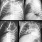
Radiographic features
Plain radiograph
Chest radiographic findings are usually subtle and non-specific. Features may include:
- apparent elevation of the cardiac apex
- right cardiac border might be indistinct due to leftward displacement and rotation of the heart
- prominent pulmonary artery segment (PA): both the medial and the lateral borders of the main PA might be seen more clearly as a result of the lack of pericardial reflection between the aorta and the PA
- lucency caused by the interposition of the lung between the aorta and main pulmonary artery segment
- the cardiophrenic space is increased on the frontal chest radiograph
- appearances may form the Snoopy sign, which is said to be pathognomonic for pericardial agenesis
CT
- may show the absence of the thin pericardial lining around the heart
- may demonstrate excessive levoposition of the heart
- the lateral and posterior portions of the pericardium may be difficult to visualize on CT
- normally, the aortopulmonary window is covered by pericardium and contains some fat. In the absence of the pericardium, lung tissue can be detected between the aorta and the main segment of the pulmonary artery.
- occasionally, there is bulging of the left atrial appendage through the defect
- may also be useful to assess for certain complication
MRI
Cardiac MRI is probably the best method for assessment.
Described features include:
- loss of direct visualization of the pericardium - low signal on T2
- levoposition of the heart
- increased cardiac mobility (e.g. excessive apical motion ) - on cine sequences
- interposition of lung tissue
- between the heart and other intrathoracic structures
- a "tongue" of lung tissue interposing between the main pulmonary artery and aorta - this is considered a very specific sign
- between the diaphragm and the base of the heart
MRI is also useful to assess for associated complications.
Treatment and prognosis
Asymptomatic isolated agenesis usually requires no treatment . Symptomatic patients who have the complete form may benefit from a pericardioplasty .
Complications
Recognized but uncommon complications include:
- cardiac valvular disease, especially insufficiency
- herniation (in cases with partial agenesis ) with incarceration of cardiac tissue, which includes atrial appendage entrapment
- compression of the left coronary artery secondary to herniation
Siehe auch:


 Assoziationen und Differentialdiagnosen zu kongenitaler Perikarddefekt: