coronal vertebral cleft

Coronal vertebral clefts refer to the presence of radiolucent vertical defects on a lateral radiograph.
Epidemiology
It is most often seen in premature male infants . As they can occur as part of normal variation (especially in the lower thoracic-upper lumbar spine of premature infants) they should not be necessarily interpreted as a malformation if seen in a newborn radiograph .
However, they can also be found in association with :
- skeletal dysplasia(s)
- chondrodysplasia punctata (all types)
- Kniest dysplasia
- mesomelic dysplasias
- metatropic dysplasia
Pathology
It often represents a delay in normal vertebral maturation and results from a failure of fusion of anterior and posterior ossification centers which remain separated by a cartilage plate.
Location
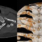
As a whole, there is a predilection for the lower thoracic and lumbar vertebral bodies .
Radiographic features
Plain radiograph
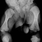
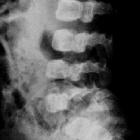
On the lateral view of the spine, it may be seen as a vertical radiolucent band just behind the midportion of the body . The affected vertebra may appear somewhat larger than those adjoining them.
Treatment and prognosis
In most cases, the vertebral clefts disappear by six months after birth .
Siehe auch:
- Schmetterlingswirbel
- angeborene Wirbelanomalien
- Chondrodysplasia punctata
- Skelettdysplasie
- Kniest-Syndrom
und weiter:


 Assoziationen und Differentialdiagnosen zu koronare Wirbelspalten: