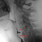
Cricopharyngeus muscle


The inferior pharyngeal constrictor muscle is one of the pharyngeal constrictor muscles. The muscle is described as having two main parts, thyropharyngeus and cricopharyngeus, which originate from the oblique line of the thyroid lamina and lateral aspect of the cricoid cartilage respectively.
Gross anatomy
The thyropharyngeus muscle arises from the (a) thyroid cartilage at the oblique line and (b) the tendinous arch that spans the cricothyroid muscle. Its fibers curve around posteriorly and insert into the midline raphe. The lower fibers are horizontal and lie edge to edge with those of the cricopharyngeus component.
The cricopharyngeus muscle is rounder and thicker than the other constrictor muscles. Its fibers run continuously from one side of the cricoid arch to the other side around the pharynx without inserting into a midline raphe posteriorly. Its muscle fibers are continuous with the circular muscular coat of the esophagus. It acts as a sphincter at the lower end of the pharynx and is always closed except for momentary relaxation during swallowing.
The cricopharyngeus has an important sphincteric function. It is physiologically in a tonic state, constricting the distal end of the pharynx (in coordination with the superior pharyngeal constrictor and the middle pharyngeal constrictor muscles). It is always closed except for momentary relaxation during swallowing. Its closure prevents air from being sucked into the upper esophagus when intrathoracic pressure falls, allowing air to be sucked only into the permanently open trachea.
Relations and/or boundaries
- The recurrent laryngeal nerve and inferior laryngeal vessels pass upwards deep to the lower border of the cricopharyngeus
- The middle and superior constrictors are enclosed by the thyropharyngeus fibers that wrap around to the midline raphe
Related pathology
The area of the pharyngeal wall at the junction between the thyropharyngeus and the cricopharyngeus is a potentially weak area. A focal weakness or deficiency in this area of the pharyngeal wall is termed a Killian dehiscence and can produce a pulsion-pseudodiverticulum, known as a pharyngeal diverticulum or pouch (Zenker diverticulum).
Summary
- origin: cricoid and thyroid cartilage
- insertion: pharyngeal raphe
- artery: ascending pharyngeal artery, branch of the external carotid artery
- nerve: pharyngeal plexus; cricopharyngeus part can also receive innervation from the recurrent laryngeal nerve and/or the external laryngeal nerve
- action: sphincteric function, swallowing
Siehe auch:
und weiter:

 Assoziationen und Differentialdiagnosen zu Musculus constrictor pharyngis inferior:
