diaphragmatic eventration


Diaphragmatic eventration refers to an abnormal contour of the diaphragmatic dome with no disruption to the diaphragmatic continuity. It typically affects only a segment of the hemidiaphragm, compared to paralysis/weakness where the entire hemidiaphragm is typically affected.
Pathology
Diaphragmatic eventration is congenital in nature and due to incomplete muscularisation of the diaphragm with a thin membranous sheet replacing normal diaphragmatic muscle. Over time this region stretches and on inspiration does not contract normally.
Causes
Congenital
Phrenic nerve agenesis.
- Failure of myotomes to migrate to a localized area in the diaphragm leading to an abnormal muscularisation.
Acquired
- Trauma/Surgery/Birth trauma leading to phrenic neve paralysis (most common cause)
Unilateral eventration may be associated with Beckwith-Weidemann Syndrome, trisomy 13, 15 or 18.
Bilateral eventration may be associated with toxoplasmosis, CMV or arthrogryposis.
Location
Congenital eventration is frequently seen in the anteromedial portion of the right hemidiaphragm, while acquired eventration is frequently seen in the left hemidiaphragm.
Radiographic features
Plain radiograph
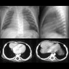
Elevation of the affected portion of the diaphragm is usually seen as a smooth hump, while the remainder of the hemidiaphragm contour is normal. The frontal X-ray may show a 'double' diaphragmatic contour, which is easily confirmed on the lateral projection.
CT
- maybe a sharp border between the eventrated portion and the remainder of the hemidiaphragm
Differential diagnosis
Possible considerations on plain film include:
- lung
- consolidation (e.g. round pneumonia)
- lung mass
- collapse
- pulmonary infarction
- pleura
- diaphragmatic mass
- diaphragmatic rupture
- Morgagni hernia
- subdiaphragmatic region
- liver/stomach mass
- subdiaphragmatic collection
- phrenic nerve palsy
- hemiplegia
Siehe auch:
und weiter:


 Assoziationen und Differentialdiagnosen zu Zwerchfellbuckel:

