hypopharyngeal squamous cell carcinoma
Squamous cell carcinoma (SCC) of the hypopharynx is relatively uncommon, carries the worst prognosis of any head and neck squamous cell carcinoma (HNSCC), and is a challenge to diagnose and treat.
Hypopharyngeal carcinoma is relatively uncommon representing only 10% of all proximal aerodigestive tract malignancies. Squamous cell carcinomas account for ~95% of all primary tumors of the hypopharynx.
Epidemiology
The epidemiology of hypopharyngeal SCC is essentially the same as that of other head and neck SCC, and is typically encountered in patients with a long history of smoking tobacco and alcohol consumption . As such they are more common in males, with perhaps the exception of posterior cricoid tumors which may be more common in women (especially of northern Europe) with Plummer-Vinson syndrome.
Clinical presentation
Most frequent presenting symptoms are those of dysphagia or odynophagia. As these tumors often present late, other frequent presenting symptoms include a neck mass (either representing the tumor or nodal metastases; the latter being the presenting symptom in up to a quarter of patients), change in voice quality (due to involvement of the larynx of laryngeal innervation), or even systemic symptoms such as weight loss .
Piriform sinus tumors have a tendency to cause referred pain to the external acoustic meatus due to cross innervation with neurons of the internal laryngeal nerve and auricular nerve, both branches of the vagus nerve (CN X) .
Stenotic tumors near the pharyngo-esophageal junction may result in severe dysphagia and even near obstruction.
Pathology
The hypopharyngeal subsite is not evenly involved :
- piriform sinus: 66-75%
- posterior pharyngeal wall
- postcricoid area/ pharyngo-esophageal junction
Risk factors
Human papillomavirus may also play a role, although it is isolated in far fewer patients with hypopharyngeal SCC (16%) compared to oropharyngeal SCC . See main HNSCC article for a general discussion.
Radiographic features
Imaging is essential not only in diagnosing likely SCC but also staging the tumor (see hypopharyngeal SCC staging).
Fluoroscopy
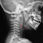
As dysphagia is a common presenting symptom, a barium swallow is often the first examination requested. Small sessile or superficially spreading lesions can be difficult or impossible to diagnose. Larger lesions may be visualized as irregular filling defects or result in asymmetry.
CT
The primary tumor typically appears as a solid soft tissue nodule or region of superficial thickening with increased enhancement. When the tumor extends beyond the confines of the pharynx, the surrounding fat planes are obliterated. It should be noted however that such stranding may be due to an inflammatory response rather than necessarily representing tumor invasion .
Careful assessment of cervical lymph nodes is essential as up to 75% of patients with hypopharyngeal SCCs have nodal metastases at the time of diagnosis .
Following irradiation, CT can be challenging, as irradiated mucosa often becomes edematous, and soft tissue fibrosis may develop, obliterating or distorting normal fat planes and potentially mimicking tumor involvement .
MRI
MRI has the ability to be superior to CT in local staging and assessing perineural spread however relatively long acquisition times and degradation by motion artifact are sometimes challenging .
- T1: intermediate to low signal mass
- T2: intermediate to high signal
- T1 C+ (GAD):
- enhancement usually present
- larger tumors of nodal metastases may be centrally necrotic
Nuclear medicine
FDG PET-CT has an increasing role play in diagnosis, staging and follow-up of head and neck malignancies, allowing identification of metabolically active tumor deposits. As is the case with FDG-PET elsewhere size is a limitation, as is movement artefact and presence of dental amalgam artefact .
Treatment and prognosis
Treatment of hypopharyngeal squamous cell carcinoma usually involves surgical resection and/or radiotherapy.
Squamous cell carcinoma of the hypopharynx carries the worst prognosis of any SCC of the upper aerodigestive tract of the head and neck both because it often presents with advanced disease. Even when prognosis is corrected for the stage, hypopharyngeal cancers continue to have poor outcomes .
- stage I-II: 47% 5-year survival
- stage III-IVb: 30% 5-year survival
- stage IVc: 16% 5-year survival
Differential diagnoses
- non-squamous cell malignancy
- accessory salivary gland tumors
- lymphoma
- radiation change in the setting of irradiation for malignancy elsewhere in the head and neck
- retropharyngeal abscess
See also
Siehe auch:
- retropharyngealer Abszess
- Plummer-Vinson-Syndrom
- hypopharyngeal SCC staging
- hypopharyngeal subsites
- SCC of the upper aerodigestive tract of the head and neck
- squamous cell carcinomas of the aerodigestive tract of the head and neck
und weiter:


 Assoziationen und Differentialdiagnosen zu squamous cell carcinoma of the hypopharynx: