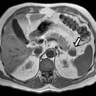
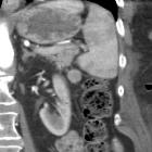
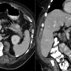
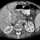
intrapancreatic splenunculus


Intrapankreatische Nebenmilzen sind kleine, in der Regel harmlose Ansammlungen von ektopem Milzgewebe, die sich während der Embryonalentwicklung außerhalb der eigentlichen Milz ansiedeln – in diesem Fall im Pankreas, typischerweise im Pankreasschwanz. Ihre Häufigkeit wird mit maximal 1 - 2 % der Nebenmilzen angegeben.
Diagnostische Bedeutung und Verwechslungsgefahr
Intrapankreatische Nebenmilzen stellen eine wichtige Differenzialdiagnose bei neoplasie- / malignomverdächtigen Raumforderungen im Pankreasschwanz dar, da sie in der Bildgebung wie ein neuroendokriner Tumor (NET) oder ein Pankreaskarzinom erscheinen können. Eine sorgfältige Bildanalyse ggf. mit mehreren Methoden sollte durchgeführt werden, um invasivere Maßnahmen zu vermeiden. Dazu gehört auch immer die Prüfung, ob evtl. ältere Aufnahmen existieren, die den Befund schon in gleicher Weise zeigen. Entsprechend der histologischen Übereinstimmung mit der normalen Milz, sollte die Nebenmilz auch im Pankreas in allen Bildgebungen mit dem Muster von normalem Milzgewebe übereinstimmen. Im Zweifel kann die definitive Diagnose durch eine Feinnadelbiopsie (EUS-FNB) oder eine Szintigrafie mit Technetium-99-markierten, hitzedenaturierten Erythrozyten gestellt werden.
Siehe auch:

 Assoziationen und Differentialdiagnosen zu intrapankreatische Nebenmilz:


