junctional parenchymal defect of kidney

Junctional
parenchymal defect of kidney • Junctional parenchymal defect of the kidney - Ganzer Fall bei Radiopaedia

Junctional
parenchymal defect of kidney • Junctional parenchymal defect of kidney - Ganzer Fall bei Radiopaedia
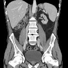
Junctional parenchymal defects in renal imaging are a normal variant, which results from the incomplete embryonic fusion of renunculi.
Radiographic features
Ultrasound
It can be seen as a triangular echogenic cortical defect, frequently seen in upper lobe parenchyma. The defect is the extension of sinus fat into the cortex, usually at the border of the upper pole and interpolar region of the kidney.
Differential diagnosis
General imaging considerations include:
- renal cortical defect
- duplex kidney
- renal angiomyolipoma (usually more round)
Siehe auch:
und weiter:


 Assoziationen und Differentialdiagnosen zu junktionaler Parenchymdefekt der Niere: