Juvenile hyaline fibromatosis
Juvenile hyaline fibromatosis is a rare autosomal recessive syndrome outlined by painful, abnormal, often deforming deposits of hyalinized fibrous material in the extracellular matrix of the skin, subcutaneous soft tissues and gums. It is listed under fibroblastic and myofibroblastic tumors in the WHO classification of soft tissue tumors.
Terminology
Alternative terms for juvenile hyaline fibromatosis include hyaline fibromatosis syndrome or infantile systemic hyalinosis .
Epidemiology
Juvenile hyaline fibromatosis is an extremely rare disorder typically occurring in infancy and childhood. It follows an autosomal recessive inheritance pattern and does not have any gender predominance .
Associations
Juvenile hyaline fibromatosis is associated with recurrent infections.
Clinical presentation
It is a progressive chronic often debilitating disease manifesting with soft tissue nodules and subcutaneous plaques or papules, gingival hypertrophy as well as other cutaneous or osseous manifestations. Systemic symptoms include failure to thrive or diarrhea .
Complications
Complications of hyaline fibromatosis include the following :
- joint contractures (very common in more than 80% )
- recurrent infections
- pericardial effusions
Pathology
Main characteristics of juvenile hyaline fibromatosis include subcutaneous nodules and gingival hypertrophy due to abundant hyaline-like deposits .
Etiology
It is an autosomal recessive disorder leading to an accumulation of collagen type VI in the extracellular matrix.
Location
Juvenile hyaline fibromatosis is a multisystem disease. Usually, the subcutaneous tissues, in particular, the scalp, the joints and the gingiva are affected. But it can also involve the myocardium, the gastrointestinal tract, lymphatic tissues as spleen and lymph nodes as well as thyroid or adrenal glands .
Macroscopic appearance
The macroscopic appearance of juvenile hyaline fibromatosis is outlined by solid, homogenous, waxy white nodules .
Microscopic appearance
The microscopic spectrum of juvenile hyaline fibromatosis lesions includes the following :
- deposits of eosinophilic non-fibrillar hyaline within the extracellular matrix
- variable number of uniform plump to spindled fibroblasts arranged in cords
- no cell atypia
- no necrosis
Immunohistochemistry
Immunohistochemistry stains can be faintly positive for vimentin or focally for smooth muscle actin.
Hyaline is diastase resistant and strongly positive for PAS, which is a special stain and not an immunostain .
Genetics
Gene mutations affect the ANTXR2 (CMG2) gene on chromosome 4q21.
Radiographic features
There are only a few reports in the literature focusing on imaging appearances of juvenile hyaline fibromatosis. The disease is characterized by subcutaneous or cutaneous nodules. Osseous involvement causes osteolytic lesions, which might be found at the skull, the long bones and phalanges of the hands and feet .
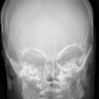
Plain radiograph
Plain radiographs might show mixed osteolytic and sclerotic lesions as well as soft tissue swelling .
MRI
Whole-body MRI might be a good imaging modality to visualize subcutaneous nodules in different locations as well as the detection of visceral involvement. However, only very few cases have been described so far .
Signal characteristics
- T1: heterogeneous, mostly isointense signal intensity
- T2: heterogeneous iso- to high signal intensity
- T1 C+ (Gd): discrete enhancement
Radiology report
The radiological report should include a description of the following:
- location and size of nodules
- relation to the muscular fascia
- relation to adjacent bones
- relationship to local nerves and vessels
- relationship to other organs
Treatment and prognosis
Management is predominantly supportive along with surgical excisions of subcutaneous nodules or gingivectomy. Recurrences are common. The main prognostic factor in juvenile hyaline fibromatosis is the age of presentation. An early presentation results in more severe disease and median survival is approximately 15 months . Mild forms might require the use of a wheelchair because of joint contractures.
History and etymology
The disease was described by Murray in 1873, who termed the disease ‘molluscum fibrosum in children’ and later by Puretic et al. in 1962, who termed it mesenchymal dysplasia. The name juvenile hyaline fibromatosis was coined by a Japanese dermatologist Yukio Kitano et al. in 1972 .
Differential diagnosis
Conditions which can mimic the appearance of juvenile hyaline fibromatosis include :
Siehe auch:


 Assoziationen und Differentialdiagnosen zu Infantile systemische Hyalinose: