lacunar infarct


Lacunar infarcts are small (<15 mm) infarcts in the distal distribution of deep penetrating vessels (lenticulostriate, thalamoperforating, and pontine perforating arteries, recurrent artery of Heubner). They result from occlusion of one of the small penetrating end arteries at the base of the brain and are due to fibrinoid degeneration.
Clinical presentation
Most lacunar infarcts are clinically silent, but repeated episodes are associated with vascular dementia. Symptomatic patients may present with lacunar stroke syndrome (LACS), one of five distinct syndromes.
Pathology
Lacunar infarcts, by definition, are caused by occlusion small penetrating end-arteries and must be smaller than 15 mm. They are thought to result primarily from in situ microatheroma formation or lipohyalinosis .
Pathologically, they are small holes of encephalomalacia and are traversed by a cob-web-like mesh of fibrous strands.
Radiographic features
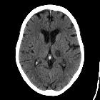
CT
In an acute setting, lacunar infarcts appear as ill-defined hypodensities.
Chronic lesions appear as hypodense foci (similar to CSF).
MRI
In an acute setting, the following signal changes are seen:
- T1: slightly hypointense
- T2/FLAIR: hyperintense
- DWI: restricted diffusion
- may demonstrate acute lesions not visible on other sequences
- T1C+: may enhance if acute (or early subacute)
Chronic lesions are isointense to CSF on all sequences but may demonstrate a peripheral T2/FLAIR hyperintense rim of marginal gliosis.
History and etymology
The term was penned by Charles Miller Fisher (1913-2012) a Canadian neurologist, who described "lacunes" (Latin: lake) of empty fluid within the brains of stroke victims post-mortem.
Differential diagnosis
- embolic stroke
- enlarged Virchow-Robin spaces
- neurocysticercosis
- choroid fissure cysts
- striatocapsular infarct
Siehe auch:
und weiter:


 Assoziationen und Differentialdiagnosen zu lacunar infarcts: