McDonald diagnostic criteria for multiple sclerosis
McDonald diagnostic criteria for multiple sclerosis are clinical, radiographic, and laboratory criteria used in the diagnosis of multiple sclerosis. They were originally introduced in 2001 and revised multiple times (see "previous versions" below) most recently in 2017 .
Criteria
The diagnosis of multiple sclerosis can be made if there is fulfillment of either of these five categories of criteria, depending on how many clinical attacks have occurred :
- ≥2 clinical attacks
- with ≥2 lesions with objective clinical evidence
- with no additional data needed
- ≥2 clinical attacks
- with 1 lesion with objective clinical evidence and a clinical history suggestive of a previous lesion
- with no additional data needed
- ≥2 clinical attacks
- with 1 lesion with objective clinical evidence and no clinical history suggestive of a previous lesion
- with dissemination in space evident on MRI
- 1 clinical attack (i.e. clinically isolated syndrome)
- with ≥2 lesions with objective clinical evidence
- with dissemination in time evident on MRI or demonstration of CSF-specific oligoclonal bands
- 1 clinical attack (i.e. clinically isolated syndrome)
- with 1 lesion with objective clinical evidence
- with dissemination in space evident on MRI
- with dissemination in time evident on MRI or demonstration of CSF-specific oligoclonal bands
Dissemination in space
Dissemination in space requires ≥1 T2-hyperintense lesions (≥3 mm in long axis), symptomatic and/or asymptomatic, that are characteristic of multiple sclerosis in two or more of the four following locations :
- periventricular (≥1 lesion, unless the patient is over the age of 50 in which case it is advised to seek a higher number of lesions)
- cortical or juxtacortical (≥1 lesion)
- infratentorial (≥1 lesion)
- spinal cord (≥1 lesion)
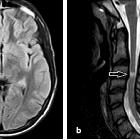
Notably, T2-hyperintense lesions of the optic nerve, such as those in a patient presenting with optic neuritis, cannot be used in fulfilling the 2017 revised McDonald criteria .
Dissemination in time
Dissemination in time can be established in one of two ways :
- a new T2-hyperintense or gadolinium-enhancing lesion when compared to a previous baseline MRI scan (irrespective of timing)
- simultaneous presence of a gadolinium-enhancing lesion and a non-enhancing T2-hyperintense lesion on any one MRI scan
Primary progressive multiple sclerosis (PPMS)
In addition to the above criteria, the McDonald criteria also define the diagnosis of primary progressive multiple sclerosis. The diagnosis now requires :
- ≥1 year of disability progression which can be determined either prospectively or retrospectively
- with two of the following:
- ≥1 T2-hyperintense lesions characteristic of multiple sclerosis in one or more of the following regions: periventricular, cortical or juxtacortical, or infratentorial
- ≥2 T2-hyperintense lesions in the spinal cord
- presence of CSF-specific oligoclonal bands
History and etymology
The criteria are named after Ian McDonald (1933-2006), a New Zealand neurologist, who devised the original criteria with his international colleagues in 2001 .
Previous versions
McDonald criteria were originally introduced in 2001 , revised in 2005 , 2010 , 2016 (by MAGNIMS) and most recently in 2017 . The 2017 revision is presented above.
Below are summaries of the previous version, that may be useful if reading earlier literature.
2001
Disseminated in space
3 out of 4 of the following:
- 1 gadolinium-enhancing lesion or 9 T2 hyperintense brain and/or cord lesions
- 1 or more infratentorial lesions
- 1 or more juxtacortical lesions
- 3 or more periventricular lesions
Disseminated in time
- 1 or more new gadolinium-enhancing lesions on a scan performed at least 3 months after onset of initial symptoms and at a new site
OR
- 1 or more new hyperintense T2 lesions compared to a scan performed at least 30 days after onset of symptoms.
These imaging criteria are combined with:
- clinical signs and symptoms
- CSF oligoclonal bands
- positive visual evoked potentials
2005
In 2005 several revisions were proposed which include
- multiple spinal lesions may be a substitute for brain and infratentorial lesion criteria, as long as they are greater than 3 mm in size, the length less than 2 vertebral body heights, and the lesion occupies only a portion of the cord cross-section
- an enhancing spinal cord lesion may be substituted for an enhancing brain lesion
- for dissemination in time, a new T2 lesion discovery interval may be reduced from 3 months to 1 month
2010
Dissemination in space
Dissemination in space requires T2 bright lesions in two or more of the following locations:
- periventricular (≥ 3 lesions)
- cortical or juxtacortical (≥1 lesion)
- optic nerve (≥1 lesion)
- infratentorial (≥1 lesion)
- spinal cord (≥1 lesion)
Dissemination in time
Dissemination in time can be established in one of two ways:
- a new lesion when compared to a previous scan (irrespective of timing)
- T2 bright lesion and/or gadolinium-enhancing
- presence of an asymptomatic enhancing lesion and a non-enhancing T2 bright lesion on any one scan
Primary progressive multiple sclerosis (PPMS)
In addition to the above criteria, the diagnosis of primary progressive multiple sclerosis has also been revised. The diagnosis now requires:
- ≥1 year of disease progression (this can be determined either prospectively or retrospectively)
- fulfill the general criteria for dissemination in space
2017
As in previous iterations of these criteria, the diagnosis of multiple sclerosis requires clinical and radiographic evidence . The two major changes in the 2017 revision are :
- the early diagnosis of multiple sclerosis can be made in patients with clinically isolated syndrome, demonstration of dissemination of space on MRI, and the presence of CSF-specific oligoclonal bands, without the need for demonstration of dissemination of time on MRI
- symptomatic and/or asymptomatic MRI lesions, except those in the optic nerve, can be considered in the determination of dissemination in space or time
Siehe auch:
- Encephalomyelitis disseminata
- McDonald diagnostic criteria 2001 - 2005
- MAGNIMS consensus on MRI diagnosis of multiple sclerosis
- McDonald diagnostic criteria 2010
und weiter:


 Assoziationen und Differentialdiagnosen zu McDonald Diagnostic Criteria for MS: