miliary tuberculosis

Disseminated
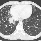
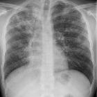
tuberculosis after anti-TNFα therapy for Crohn\"s disease. At admission radiographs showed the appearance of a \"hazy\" micronodular pattern symmetrically involving both lungs, without signs of pleural effusion.

Miliary
tuberculosis • Disseminated tuberculosis - Ganzer Fall bei Radiopaedia

Toddler with
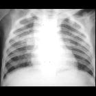
fever. CXR AP shows innumerable small pulmonary nodules that are uniformly distributed throughout the lungs.The diagnosis was miliary tuberculosis pneumonia.

Miliary
tuberculosis • Tuberculosis - Ganzer Fall bei Radiopaedia

Miliary
tuberculosis • Disseminated tuberculosis with spondylitis (Pott disease) and massive iliopsoas abscesses - Ganzer Fall bei Radiopaedia

Miliary
tuberculosis • Miliary tuberculosis - Ganzer Fall bei Radiopaedia

Miliary
tuberculosis • Miliary tuberculosis - Ganzer Fall bei Radiopaedia

Miliary
tuberculosis • Miliary tuberculosis - Ganzer Fall bei Radiopaedia

Miliary
tuberculosis • Miliary tuberculosis - Ganzer Fall bei Radiopaedia

Miliary
tuberculosis • Miliary tuberculosis - Ganzer Fall bei Radiopaedia

Miliary
tuberculosis • Tuberculosis - CNS and chest - Ganzer Fall bei Radiopaedia

Miliary
tuberculosis • Miliary tuberculosis - Ganzer Fall bei Radiopaedia

Miliary
tuberculosis • Miliary tuberculosis - Ganzer Fall bei Radiopaedia

Extrapulmonary
tuberculosıs: an old but resurgent problem. Radiologic images of a 38-year-old-female who has a diagnosis of miliary TB. Multiple tuberculomas (red arrows) show peripheral rim-like enhancement on post-contrast T1 weighted image in the cerebellum (a). On T2-weighted images (b, c) demonstrate focal lesions (tuberculomas) with central isointense signal and peripheral hyperintensity in gray matter (blue arrows). Multiple nodules secondary to miliary TB are seen (d)


Floride,
kavernisierende Tuberkulose. Vor allem rechts basal auch miliare Herde.

Floride,
kavernisierende Tuberkulose. Vor allem rechts basal auch miliare Herde.

Floride,
kavernisierende Tuberkulose. Vor allem rechts basal auch miliare Herde.


Preschooler
with cough and fever. CXR AP shows right hilar lymphadenopathy and innumerable small pulmonary nodules that are uniformly distributed throughout the lungs.The diagnosis was miliary tuberculosis pneumonia.

Miliary
tuberculosis • Miliary tuberculosis - Ganzer Fall bei Radiopaedia
Miliary tuberculosis is an uncommon pulmonary manifestation of tuberculosis. It represents haematogenous dissemination of uncontrolled tuberculous infection and carries a relatively poor prognosis. It is seen both in primary and post-primary tuberculosis and may be associated with tuberculous infection in numerous other tissues and organs.
Radiographic features
Plain radiograph
Miliary deposits appear as 1-3 mm diameter nodules, which are uniform in size and uniformly distributed.
CT
Similar findings to plain radiograph but may more elaborately show extent and distribution.
Treatment and prognosis
If treatment is successful no residual abnormality remains.
See also
Siehe auch:
und weiter:

 Assoziationen und Differentialdiagnosen zu Miliartuberkulose:

