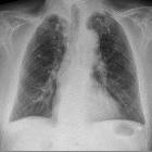
verkalkte Lungenherde

CT findings
of high-attenuation pulmonary abnormalities. Metastatic pulmonary calcification in a 24-year-old man who had known chronic renal failure. Axial CT at a mediastinal and b parenchymal windows demonstrates bilateral centrilobular fluffy ground-glass nodular opacities that contain foci of calcification (arrows)

Metabolic and
storage lung diseases: spectrum of imaging appearances. A 54-year-old man with fibrosing mediastinitis, mitral valve stenosis and nodular pulmonary ossification. a Unenhanced HRCT image shows high attenuation ground-glass opacities in the right upper and lower lobes with punctate calcifications (arrowheads). b Unenhanced HRCT image shows soft tissue thickening and dense calcification (arrow) at the confluence of the right pulmonary veins

Calcified
pulmonary nodules • Nodular pulmonary amyloidosis - Ganzer Fall bei Radiopaedia

Calcified
pulmonary nodules • Calciphylaxis and metastatic pulmonary calcification - Ganzer Fall bei Radiopaedia

Calcified
pulmonary nodules • Silicosis with progressive massive fibrosis - Ganzer Fall bei Radiopaedia

Calcified
pulmonary nodules • Calcified pulmonary metastases - Ganzer Fall bei Radiopaedia

Calcified
pulmonary nodules • Alveolar microlithiasis - Ganzer Fall bei Radiopaedia

Varicella
pneumonia • Healed varicella pneumonia - miliary opacities - Ganzer Fall bei Radiopaedia

Calcified
pulmonary nodules • Miliary tuberculosis - Ganzer Fall bei Radiopaedia

Metabolic and
storage lung diseases: spectrum of imaging appearances. A 54-year-old man with nodular parenchymal amyloidosis. a Coronal reformatted HRCT image shows scattered punctate calcifications (arrowheads) in both lungs with patchy consolidation (arrows) in the right lung. In addition, a moderate right pleural effusion is noted. b Axial HRCT image shows punctate calcifications (arrowheads) in the lungs, a small right pleural effusion, and right paratracheal lymphadenopathy (arrow)

CT findings
of high-attenuation pulmonary abnormalities. Lung carcinoma. Axial CT shows eccentric calcification in a malignant mass, invading the mediastinum and right hilar region (arrows)

Hamartom der
Lunge; nicht histologisch bestätigt, jedoch typischer Befund: glatt begrenzter, über lange Zeit konstanter Befund mit Verkalkung.

CT findings
of high-attenuation pulmonary abnormalities. Hamartomas. Axial CT shows popcorn calcification in a benign solitary pulmonary nodule (arrow)

CT findings
of high-attenuation pulmonary abnormalities. Pulmonary alveolar microlithiasis. Axial CT at a mediastinal and b parenchymal windows shows diffuse numerous dense micronodules with calcified thickening of interlobular septa and subpleural cysts

CT findings
of high-attenuation pulmonary abnormalities. Silicosis. Axial CT at the mediastinal window shows multiple calcified nodules with a conglomerate mass of fibrosis in the upper lobes (arrows)

Zoledronic
acid in metastatic chondrosarcoma and advanced sacrum chordoma: two case reports. a Thoracic CT scan in the patient with chondrosarcoma shows at right the lesion involving muscles and ribs. Lung metastases were visualized. b Coronal section displays the large tumor.

Pulmonary
epithelioid hemangioendothelioma accompanied by bilateral multiple calcified nodules in lung. Radiographs of chest. Chest radiograph (A) and computerized tomograph (B) show an approximately 6.0 cm sized well-demarcated mass in the right lung and multiple calcified small nodules in both lungs.

Dendriform
pulmonary ossification in a case of usual interstitial pneumonia. Axial chest CT MIP image shows subpleural calcifications with branching and reticular appearance in the right lung. Other similar calcifications, less extensive, are present in the left lung.

Carney triad
(ECR 2016 Case of the Day). Coronal CT image shows same findings. These lesions should not be confused with calcified metastases.

Carney triad
(ECR 2016 Case of the Day). Follow-up CT 6 years later shows further calcification and enlargement of prior lung lesions, as well as appearance of a new nodule in the left lower lobe (arrow).

Carney triad
(ECR 2016 Case of the Day). Chest CT scan shows bilateral calcified lung nodules suggesting pulmonary chondromas.

Calcified
pulmonary nodules • Papillary thyroid carcinoma with miliary metastases - Ganzer Fall bei Radiopaedia

Acute
pneumonia evolution. MIP and coronal reconstruction with multiple small calcified nodules of chronic varicella pneumonia.
Calcified pulmonary nodules are a subset of hyperdense pulmonary nodules and a group of nodules with a relatively narrow differential.
Pathology
Etiology
The most common cause of nodule calcification is granuloma formation, usually in the response to healed infection.
- healed infection
- calcified granulomata, e.g. thoracic histoplasmosis, recovered miliary tuberculosis (rare)
- most common
- 2-5 mm
- calcification may be central or diffuse
- usually with calcification of hilar/mediastinal nodes
- healed varicella pneumonia
- micronodular (1-3 mm)
- no associated nodal calcification
- calcified granulomata, e.g. thoracic histoplasmosis, recovered miliary tuberculosis (rare)
- occupational disease/pneumoconioses
- silicosis
- associated with nodal egg-shell calcification
- multiple small densely calcified nodules in mid and upper zones
- coalworker's pneumoconiosis
- smaller nodules which may not be visible on plain film
- associated with minimal symptoms
- silicosis
- pulmonary hamartomas
- metastatic pulmonary calcification
- typically nodules are poorly defined and larger (3-10 mm)
- calcium and phosphate metabolism abnormalities
- chronic renal failure
- multiple myeloma
- secondary hyperparathyroidism
- massive osteolysis caused by metastases
- intravenous calcium therapy
- pulmonary hemosiderosis
- idiopathic pulmonary hemosiderosis
- recurrent alveolar hemorrhage
- centrilobular nodular opacities
- mitral stenosis
- small multifocal calcified nodules
- Goodpasture syndrome
- idiopathic pulmonary hemosiderosis
- pulmonary alveolar microlithiasis
- tiny micronodules
- apparent calcification of interlobular septa
- small subpleural cysts
- sarcoidosis
- calcifying fibrous pseudotumor of lung
- pulmonary amyloidosis
- pulmonary hyalinising granuloma
- calcified pulmonary metastases
See also
- pulmonary calcification
- pulmonary nodules
- hyperdense pulmonary nodules
- calcified pulmonary nodules
- non-calcified hyperdense pulmonary nodules
- hyperdense pulmonary nodules
- hyperattenuating pulmonary abnormalities
- differential diagnosis of calcific pulmonary densities
Siehe auch:
- Sarkoidose
- Osteosarkom
- postspezifische Granulome der Lunge
- Silikose
- Multiples Myelom
- Lungenrundherd
- Miliartuberkulose
- pulmonales Hamartom
- Pneumonokoniose
- multiple kleine hyperdense Lungenherde
- Hämosiderose der Lunge
- Goodpasture-Syndrom
- Mitralklappenstenose
- alveoläre Mikrolithiasis
- Eierschalenverkalkungen
- verkalkte Lungenmetastasen
- nicht verkalkte hyperdense Lungenherde
- Varizellenpneumonie
- pulmonale Calciphylaxie
- granulomatous lung disease
- pulmonale Verkalkungen
- Verkalkungen Tuberkulose
- Lungenmetastasen bei Osteosarkom
- pulmonales Granulom
- noduläre pulmonale Ossifikation
- diffuse Lungenverkalkungen
- hyperdense pulmonale Raumforderungen
- coalworker's pneuomconiosis
und weiter:

 Assoziationen und Differentialdiagnosen zu verkalkte Lungenherde:
hyperdense
pulmonale Raumforderungen


