Pneumonokoniose


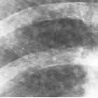
Röntgenbild
einer Silikose mit multiplen kleinnodulären Verdichtungen. Zusätzlich alte Rippenfrakturen links.


Chest X-ray
in asbestosis shows plaques above diaphragm Source: Early Asbestosis in a Retired Pipe Fitter http://clinicalcases.blogspot.com/2004/03/early-asbestosis-in-retired-pipe.html Clinical_Cases: I made the photo myself, licensed under Creative Commons license.

Pneumoconiosis
• Calcified pleural plaques from asbestos exposure - Ganzer Fall bei Radiopaedia

Pneumoconiosis
• Mixed pneumoconiosis - silicosis with PMF and asbestosis - Ganzer Fall bei Radiopaedia

Pneumoconiosis
• Silicosis with progressive massive fibrosis - Ganzer Fall bei Radiopaedia

Imaging
diagnosis of classical and new pneumoconiosis: predominant reticular HRCT pattern. Subpleural dots and subpleural lines in asbestosis. Some dots are located a few millimeters from the pleura (arrows). In asbestosis, subpleural line is much closer to the pleural surface and the distance of the subpleural lines from the inner chest wall is 2 to 3 mm (arrowheads). The subpleural lines look like connected subpleural dots arranged along the inner chest wall

Imaging
diagnosis of classical and new pneumoconiosis: predominant reticular HRCT pattern. 61-year-old man with inhalational talc pneumoconiosis employed in talc industry for 20 years. a HRCT scan shows diffusely distributed small nodules. They are separated from the pulmonary vein or the pleura at a distance of about 2–3 mm and are separated regularly from each other at a distance of about 2–3 mm. b CT scan at mediastinal setting of 61-year-old man with inhalational talc pneumoconiosis shows large opacity and lymph nodes containing high-attenuation material

Imaging
diagnosis of classical and new pneumoconiosis: predominant reticular HRCT pattern. HRCT scans of pulmonary aluminosis. a 58-year-old man with pulmonary aluminosis. HRCT scan of pulmonary aluminosis mimicking sarcoidosis. Ground-glass opacities, small nodular opacities, and traction bronchiectasis are seen predominantly around the bronchovascular bundles. Nodules are located in both centrilobular and paralobular regions. b 52-year-old man with pulmonary aluminosis. HRCT shows traction bronchiectasis and ground-glass opacity predominantly in the upper lungs. Multiple bullae and centrilobular nodules are also seen

Imaging
diagnosis of classical and new pneumoconiosis: predominant reticular HRCT pattern. HRCT scans of heard metal pneumoconiosis. a 62-year-old man with hard metal pneumoconiosis. Upper-lung predominant fibrosis in hard metal pneumoconiosis. HRCT scan shows prominent interstitial thickening, irregular peribronchovascular thickening, and traction bronchiectasis in upper lung zones. b 32-year-old man with hard metal pneumoconiosis. Early stage of hard metal pneumoconiosis. HRCT scan shows ground-glass opacities and centrilobular nodules predominantly in the peripheral portions. c 53-year-old man with hard metal pneumoconiosis. HRCT scan shows patchy and irregular ground-glass opacity and traction bronchiectasis diffusely distributed in the lung. Bullae are seen in the subpleural region.

Imaging
diagnosis of classical and new pneumoconiosis: predominant reticular HRCT pattern. 59-year-old man with hard metal pneumoconiosis. Initial HRCT scan (a) shows scattered areas of ground-glass attenuation associated with fine reticulation and mild traction bronchiectasis. HRCT obtained 3 years later (b) reveals increase in parenchymal abnormalities in extent, prominent reticulation, and progression of traction bronchiectasis. HRCT obtained 5 years later (c) shows dense increased parenchymal opacities with traction bronchiectasis

Imaging
diagnosis of classical and new pneumoconiosis: predominant reticular HRCT pattern. HRCT scan of a 34-year-old man with indium lung. Tiny centrilobular nodules (arrows) are scattered in both lungs
Pneumoconioses are a broad group of lung diseases that result from inhalation of dust particles. It is therefore considered part of the spectrum of inhalational lung disease, and also occupational lung diseases.
Pathology
Etiology
The offending agents are mainly mineral dust. They can be broadly classified as :
- fibrotic
- non-fibrotic
Associations
Siehe auch:
- Silikose
- Asbestose
- Caplan-Syndrom
- Siderose
- Talkosis
- stannosis
- giant cell interstitial pneumonia
- byssinosis
- coal workers pneumoconiosis (CWP)
- Aluminose
- baritosis
- Berylliose
- hard-metal pneumoconiosis
und weiter:
- verkalkte Lungenherde
- Kerley-Linien
- Pneumocystis jiroveci Pneumonie
- miliare Lungenherde
- Interstitielle Lungenerkrankung
- causes of pulmonary arterial hypertension
- Hämosiderose der Lunge
- interstitial pneumonia
- Anthrakose
- miliary nodules in the exam
- hard metal pneumoconiosis
- pulmonary stannosis
- excipient lung disease
- progressive massive Fibrose
- nicht verkalkte hyperdense Lungenherde
- Pulmonary talc granulomatosis
- pulmonary baritosis
- inorganic dust
- Siderofibrose
- CT/HRCT - Klassifikation ICOERD
- Papierstaub Lunge

 Assoziationen und Differentialdiagnosen zu Pneumonokoniose:


