coal workers' pneumoconiosis
Coal workers' pneumoconiosis (CWP) is an occupational disease (type of pneumoconiosis) caused by exposure to coal dust free of silica (washed coal). Histologically, CWP is classified according to disease severity into simple (presence of coal macules) and complicated (with progressive massive fibrosis) .
Pathology
There are three main effects from carbon (inclusive of coal dust) on the lungs:
- anthracosis: relatively harmless deposition of carbon dust
- simple coal worker's pneumoconiosis (CWP)
- complicated coal worker's pneumoconiosis (progressive massive fibrosis): occurs in about of 10% of CWP and requires several years to develop
Along with silicosis, asbestosis, berylliosis, and talcosis, CWP is a fibrotic pneumoconiosis.
Radiographic features
Despite histologic differences between silicosis and CWP, the two entities are not easily distinguished from one another on imaging studies.
Simple CWP
Plain radiograph
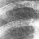
Simple CWP appears as small (1-5 mm) round nodular opacities with an occasional appearance of reticular or reticulonodular opacities. Compared to nodules in silicosis, the nodules in CWP tend to have less well-defined margins and a more granular appearance.
Between 10-20% of patients with simple CWP have small central calcifications within these nodules on chest radiograph, which are contrasted to the more diffuse calcifications of silicosis . The eggshell calcification pattern pathognomonic of simple silicosis is seen in less than 2% of patients with simple CWP .
CT
There is often a diffuse (typically perilymphatic) distribution of small nodules throughout the lungs, which tend to favor the upper lungs. About 30% of patients have calcifications with nodules on CT. Hilar or mediastinal lymph node enlargement has also been reported in 30% of patients .
Complicated CWP
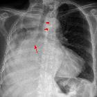
Plain radiograph
Large opacities representing progressive massive fibrosis may be seen in complicated CWP. These fibrotic masses may either have irregular borders with associated surrounding emphysema, or regular borders without surrounding emphysema .
Lung cancer may present with a similar appearance, and differentiation is obviously important.
MRI
Compared with the signal intensity of muscle, complicated CWP appears as a low signal intensity lesion on both T1 and T2 weighted images. This is in contrast to neoplastic processes that have high signal intensity on T2 weighted images . On contrast administration, complicated CWP lesions frequently demonstrate peripheral enhancement .
Nuclear medicine
The role of PET-CT in the diagnosis of malignancy in the setting of pneumoconiosis remains unclear, as both lung cancer and the fibrotic mass of complicated CWP may demonstrate increased uptake of FDG .
Complications
- increased risk of tuberculosis
- development of chronic obstructive airways disease
History and etymology
The first published case report on CWP was by Gregory (1831) in a British coal miner. For a period of time, the similarities on chest radiograph between silicosis and CWP lead to the hypothesis that CWP was a variant of silicosis. Further pathologic analysis has established CWP as a separate entity .
Differential diagnosis
General imaging differential considerations include:
Siehe auch:
- Tuberkulose
- Lungenkarzinom
- Silikose
- Chronisch obstruktive Lungenerkrankung
- Pneumonokoniose
- Asbestose
- Talkosis
- Eierschalenverkalkungen
- progressive massive Fibrose
- perilymphatic distribution of small nodules
und weiter:


 Assoziationen und Differentialdiagnosen zu coal workers pneumoconiosis (CWP):