Neuroakanthozytose

Neuroacanthocytosis syndromes (NAS), previously known collectively as Levine-Critchley syndrome, are characterized by basal ganglia degeneration, acanthocytosis, and normal serum lipoprotein.
There are four core NAS:
- chorea-acanthocytosis (ChAc)
- McLeod syndrome (MLS)
- Huntington disease-like 2 (HDL2)
- pantothenate kinase-associated neurodegeneration (PKAN)
Epidemiology
NAS are incredibly rare, with an estimated prevalence of 1-5:1000000 .
Clinical presentation
The presentation within and between each NAS is heterogeneous. Generally, patients present with movement disorder (commonly chorea and dystonia), cognitive decline and psychiatric disturbance .
Acanthocytosis, red blood cells defined by the presence of multiple thorny projections, is most often seen in ChAc and MLS, and less frequently in HDL2 and PKAN. The presence of acanthocytes is not necessary for diagnosis .
Elevated creatinine kinase is often found in ChAc and MLS.
Unique features of chorea-acanthocytosis
Of note, the finding of 'feeding dystonia' is pathognomonic of ChAc. Upon food touching the tongue, the tongue protrudes and forces the bolus out. In addition, the presence of 'rubber-man appearance', truncal instability and loss of axial tone with sudden spasms of flexion and extension, is highly suggestive of ChAc .
Pathology
Etiology
Chorea-acanthocytosis
ChAc is caused by mutations in the gene encoding chorein, vacuolar protein sorting 13 homolog A (VPS13A) . The function of chorein is unclear, but has been shown to be of importance in exocytosis and cytoskeleton structure .
McLeod syndrome
The McLeod blood group disorder arises from X-linked mutations in the XK gene at the Xp21.1 locus. The XK locus controls the expression of the ubiquitously expressed XK protein, which is predominantly found in nervous tissue, the myocardium and the erythrocytes cell surface . XK forms a disulphide bond with erythrocyte surface Kell antigens, forming a complex. XK and Kell expression is thereby correlated, and so reduced XK causes a reciprocal reduction in Kell. Most carriers of the McLeod blood group will develop the central nervous system features of MLS in later life. Of note, due to myocardial XK protein expression, cardiomyopathy often occurs in MLS. Upon laboratory testing, diminished Kell antigen expression should be seen on the red cell surface of MLS patients .
Huntington disease-like 2
Trinucleotide repeat expansions of junctophilin 3 (JPH3) are found in HDL2, which are thought to cause the formation of intracellular aggregates and subsequent cell death .
Pantothenate kinase-associated neurodegeneration
PKAN results from mutations in the pantothenate kinase 2 gene (PANK2) at the 20p13 locus. PANK2 encodes an enzyme, pantothenate kinase 2, which indirectly regulates mitochondria energy production via coenzyme A synthesis .
Summary
The genes identified in each subgroup and not clearly linked to a common pathway. However, there is a theme of membrane processing failure . It is worth noting that acanthocytes are present in a proportion of individuals affected by neurological dysfunction in other contexts. Specifically, inherited disorders of lipoprotein metabolism and in systemic diseases, such as severe malnutrition, cancer and liver cirrhosis . These are distinct from the core NAS.
Radiographic Features
MRI
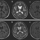
- T2/FLAIR:
- generally:
- atrophy and gliosis of the basal ganglia are typical, and so these deep brain structures may appear reduced in size
- regions of the basal ganglia may be hyperintense, indicating gliosis
- specifically:
- the head of the caudate is most often affected in ChAc, with the putamen, globus pallidus and substantia nigra affected to a lesser extent
- bilateral atrophy of the caudate and putamen is again observed in MLS and HDL2
- PKAN is characterized by an eye of the tiger iron deposition pattern - a round hyperintensity in the anteromedial globus pallidus
- generally:
Treatment and prognosis
There are no curative therapies for NAS, with clinical management focusing on symptom control and quality of life .
History and etymology
Acanthos is the Greek word for "thorn", from which the name of the red blood cell abnormality in NAS, acanthocytosis, is derived. The first case reports of adult acanthocytosis associated with neurological dysfunction, by MacDonald Critchley and Irvine Levine, date to the late 1960s . Interesting, NAS was originally known as Levine-Critchley syndrome, however, modern techniques could not confirm the originally identified families had NAS, as they were lost to follow-up, thus the eponymous term has been abandoned .
Siehe auch:
und weiter:


 Assoziationen und Differentialdiagnosen zu Neuroakanthozytose: