pancreatic islet cell tumours


Insulinomas are the most common sporadic endocrine tumor of the pancreas.
On imaging, they usually present as small well-defined hypervascular tumors that may be found anywhere in the pancreas.
Epidemiology
Account for 40% of syndromic pancreatic endocrine tumors. The overall incidence is of ~0.0003%.
Clinical presentation
Typically insulinomas present with Whipple's triad consisting of:
As with other endocrine tumors of the pancreas, there is an association with multiple endocrine neoplasia type I (MEN I).
Pathology
They develop from ductal pluripotent cells into unregulated cells secreting insulin. The beta cells of the islets of Langerhans normally secrete insulin. Approximately 10% of insulinomas are multiple and 10% malignant.
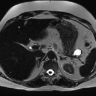
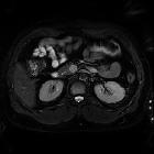
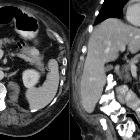
Radiographic features
These tumors can be relatively small and multiphase contrast-enhanced thin slice cross-sectional imaging is ideal. Most insulinomas are small (90% are <2 cm at presentation ) and hypervascular. They may contain calcifications. Malignant tumors tend to be larger. Equally distributed between the head, body, and tail of the pancreas.
CT
They tend to be hyperattenuating on arterial phase and, therefore, dedicated protocols with arterial or pancreatic phase imaging may aid in better detection. Some may show calcification.
MRI
Dynamic MRI with fast gradient echo sequences following a bolus injection of contrast medium may aid in the detection of these tumors :
- T1 C+ (Gd): typically shows enhancement, although contrast enhancement may not improve tumor visualization compared with non-contrast images
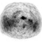
Nuclear medicine
Ga-68 DOTATATE PET-CT
About 80% of insulinomas express the somatostatin receptors 2, and the Ga-68 DOTATATE scans have a high affinity for these receptors and, therefore, have high sensitivity in the detection of these tumors, particularly for low-grade and well-differentiated ones . The sensitivity of this study has been reported in up to 90% , when assessing insulinomas specifically, and ranging between 90-100% for pancreatic neuroendocrine tumors as whole .
PET-CT is also useful in excluding additional pancreatic neuroendocrine tumors eventually not detected on CT or MRI, particularly, in inherited syndromes such as MEN1 .
F-18 FDG PET-CT
Neuroendocrine tumors are slow-growing tumors that usually have slow metabolic activity in their initial stages and, therefore, are not notably avid on F-18 FDG PET-CT . Tumors with a higher grade or poorly differentiated tend to show marked uptake .
Siehe auch:
- Pankreastumoren
- Gastrinom
- Hypervaskularisierte Pankreasläsionen
- Neuroendokriner Tumor
- multiple endokrine Neoplasie Typ 1
- neuroendokrine Pankreastumoren
- solide Pankreastumoren
- Glukagonom
- Somatostatin-Rezeptor-Szintigrafie
- benigne Pankreastumoren
- Whipple-Trias
- Somatostatinom
- Pankreasmetastasen (Nierenzellkarzinom)
- VIPom
- zystischer neuroendokriner Tumor des Pankreas
- Inselzellkarzinom
und weiter:


 Assoziationen und Differentialdiagnosen zu Insulinom:


