pelvine Lipomatose


Pelvic lipomatosis or pelvic fibrolipomatosis represents excessive deposition of fat in pelvis due to overgrowth of adipose cells leading to compression of pelvic organs.
Epidemiology
The condition usually presents in patients 20-50 years of age. The condition is predominantly (~66% of cases) seen in African-Americans.
Clinical presentation
Patients usually present with dysuria, hematuria, urgency, urinary incontinence, and/or constipation due to compression of the genitourinary system, vascular system, and lower gastrointestinal tract.
Radiographic features
Plain radiograph
Findings are non-specific and non-diagnostic with areas of lucency around the bladder.
IVP
Pear shaped or inverted teardrop bladder with dilated ureter.
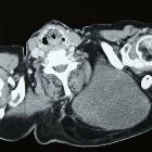
CT
CT findings are diagnostic with areas of symmetric fat density in the pelvic region. Pear-shaped bladder can be seen. The sigmoid may be stretched and surrounded by excess pelvic fat. It also can extend to the abdomen with peripheral displacement of bowel loops.
MRI
Similar to CT findings with hyperintensity noted on T1 in areas containing fat and iso to hyperintensity noted on T2W imaging.
Differential diagnosis
The differential is mainly that of inflammatory conditions resulting in fatty infiltration or other fatty masses.
- proctitis
- differentiated by a history of radiotherapy to the pelvis for proctitis
- ulcerative colitis
- lipoma or liposarcoma
Siehe auch:
und weiter:


 Assoziationen und Differentialdiagnosen zu pelvine Lipomatose: