persistent stapedial artery


The persistent stapedial artery (PSA) is an abnormal small vessel arising from the petrous portion of the internal carotid artery and crossing through the middle ear. It results from the failure of regression of the embryonic stapedial artery.
Epidemiology
The prevalence is thought to range from 0.02 to 0.48% in the population.
Clinical presentation
- pulsatile tinnitus
- conductive hearing loss due to ankylosis of the stapes
- sensorineural hearing loss due to an erosion of otic capsule (rare)
- mass seen over the promontory in the middle ear while performing an otoscopic examination
- incidental finding during surgery that can lead to hemorrhage if not properly identified
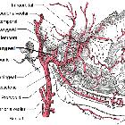
Gross anatomy
- arises from the proximal hyoid artery at 4-5 weeks of life
- passes through the obturator foramen of stapes
- branches into upper and lower divisions
- posterior division of the upper branch becomes middle meningeal artery
- lower branch (maxillomandibular) has two branches
- mandibular artery (becomes inferior alveolar artery in adult)
- infraorbital artery (becomes infraorbital artery in adult)
Anastomosis forms between the ventral pharyngeal artery (precursor of external carotid artery) and lower division of the stapedial artery.
As the ventral pharyngeal artery supplies flow to middle meningeal artery, stapedial artery regresses, leaving small caroticotympanic artery.
It is termed persistent as it is normally found until week 10, at which point flow reverses at the foramen spinosum which in turn induces degeneration over the course of the 3rd fetal month.
Blood supply
Can arise from:
Radiographic features
- small canaliculus originating from the petrous segment of the internal carotid artery
- linear soft tissue density crossing over the cochlear promontory
- enlarged facial nerve canal or separate canal parallel to facial nerve
- aplastic or hypoplastic foramen spinosum
- may be a normal variant or in instances where middle meningeal artery arises from the ophthalmic artery
History and etymology
It was first discovered in 1836 at postmortem examination by Austrian anatomist Joseph Hyrtl (1810-1894) , most famous for his description of the tympanomeningeal fissure.
Related pathology
- may be associated with aberrant internal carotid artery, which is formed when inferior tympanic artery anastomosis with the caroticotympanic artery; enlarged inferior tympanic canaliculus is then seen
- may be associated with other middle ear anomalies
Siehe auch:
- Arteria carotis interna
- Nervus facialis
- Stapes
- aberrante Arteria carotis interna
- absent foramen spinosum
- Arteria meningea media
- Rami caroticotympanici
und weiter:


 Assoziationen und Differentialdiagnosen zu persistierende Arteria stapedia: