Arteria meningea media


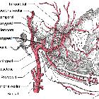
The middle meningeal artery is the dominant supply of the cranial dura. It arises from the first part of the maxillary artery, a terminal branch of the external carotid artery. It enters the middle cranial fossa via the foramen spinosum. Here it gives off two basal branches—the petrosal branch and the cavernous branch—before dividing into anterior and posterior divisions :
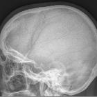
- The anterior division runs anterolaterally through the middle cranial fossa on the greater wing of sphenoid before coursing superiorly, often grooving the bone, and passes under the pterion before giving its terminal branches over the upper parietal bone.
- The posterior division runs horizontally posteriorly over the squamous part of the temporal bone to give rise to its terminal branches over the lower parietal bone.
Gross anatomy
Relations and/or Boundaries
In its extracranial portion, the middle meningeal artery runs vertically through the roots of the auriculotemporal nerve.
Variant anatomy
The ophthalmic artery infrequently arises from the middle meningeal artery .
Conversely, the middle meningeal artery infrequently arises from branches of the internal carotid artery. The most common of these origins is the ophthalmic artery, when it may be termed the "ophthalmic-middle meningeal artery" , or an ophthalmic branch like the lacrimal artery . Rarely, the middle meningeal artery arises from a persistent stapedial artery from the petrous segment of the internal carotid artery, when it may be termed the "stapedial-middle meningeal artery" .
Rarely, the middle meningeal artery has anastomoses with or origin from the basilar artery .
Related pathology
- epidural hematoma, which is most commonly due to traumatic rupture of the middle meningeal artery
- chronic recurrent subdural hematoma, which may be treated with middle meningeal artery embolization
- intracranial dural arteriovenous fistula, for which the middle meningeal artery is the most common arterial feeder
- meningioma, which may be embolized preoperatively via the middle meningeal artery
Siehe auch:
und weiter:

