pleomorphic liposarcoma


Pleomorphic liposarcomas are high-grade malignant neoplasms arising from adipocytic tissues characterized by a variable number of pleomorphic lipoblasts and the absence of areas of well-differentiated liposarcoma and other lines of differentiation.
Epidemiology
Pleomorphic sarcomas are rare and make up for less than 5% of all liposarcomas and for about 20% of pleomorphic sarcomas. Most commonly they occur in the 7 decade of life with men slightly more frequently affected. In children, they are extremely rare .
Clinical presentation
The most frequent symptom is that of a fast-growing painless tumor, pain and/or symptoms related to organ displacement are less common complaints .
Pathology
Pleomorphic liposarcomas are highly malignant neoplasms characterized by pleomorphic lipoblasts with features of pleomorphic spindle cell sarcoma or myxofibrosarcoma and in the case of the epithelioid subtype with sheets of carcinoma-like epithelioid cells . They commonly have necrotic and myxoid changes .
Location
The most frequent sites of involvement by pleomorphic liposarcomas are the extremities in about ⅔ of the cases. The lower limbs are more commonly affected. Less frequent locations include the following :
- trunk: thoracic and abdominal wall
- retroperitoneum
- spermatic cord
Rare sites of involvement are :
Subtypes
There is a single subtype, the epithelioid liposarcoma.
Macroscopic appearance
Macroscopically pleomorphic liposarcomas are usually large when detected, not encapsulated, sometimes well-demarcated or multinodular other times ill-defined and infiltrative and of yellowish-white color. They often feature areas of necrosis or myxoid change .
Microscopic appearance
The microscopic appearance of pleomorphic liposarcomas includes the following features :
- infiltrative margins
- varying proportions of pleomorphic lipoblasts
- non-lipogenic background of undifferentiated pleomorphic sarcoma with spindle cells and multinucleated giant cells
- possibly myxofibroma-like areas (≈50%)
- areas of necrosis (≈50%)
- epithelioid morphology (≈25%)
Immunohistochemistry
Different than well-differentiated or dedifferentiated liposarcoma immunohistochemistry stains are usually negative for MDM2 and/or CDK4. The epithelioid subtype might show positive stains for melan-A and/or keratins .
Genetics
Genetically most mutations involve TP53 and/or NF1. No amplification involving the MDM2 nuclear gene is seen .
Radiographic features
Pleomorphic liposarcomas look like nonspecific soft tissue masses with heterogeneous areas of necrosis and hemorrhage .Compared to other liposarcomas they contain less or no fatty-tissue. Like other tumors, they displace other organs or tissue.
Ultrasound
Usually appears as multilobulated with areas of low echogenicity .
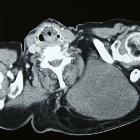
CT
CT usually shows an unspecific heterogeneous soft- tissue density mass isodense to muscle with streaky enhancing areas .
MRI
On MRI pleomorphic liposarcomas will display a soft tissue mass with fibrous components that enhance .
- T1: heterogeneous hypointense
- T2: heterogeneous hyperintense similar to or slightly lower than fat
- T1 C+ (Gd): heterogeneous enhancement
Radiology report
The radiological report should include a description of the following:
- form, location and size
- tumor margins
- amount of solid and lipoid components
- relation to other organs and/or structures
Treatment and prognosis
The management of pleomorphic liposarcomas includes resection and also shows a response to the combination of ifosfamide and doxorubicin, which makes (neo-) adjuvant chemotherapy another option . Local recurrence happens in about 30-50%, distant metastases occur in up to 50% of the cases and the 5-year survival is in the range of 60% .
Increased tumor depth and more central location, as well as a greater size and higher mitotic activity, are associated with worse outcome .
Differential diagnosis
Conditions which can mimic the clinical or radiological presentation of pleomorphic sarcoma are any fast-growing soft tissue tumors and include :
- myxoid pleomorphic liposarcoma
- myxoid liposarcoma
- dedifferentiated liposarcoma
- fibrosarcoma
- myxofibrosarcoma
See also
Siehe auch:
und weiter:


 Assoziationen und Differentialdiagnosen zu pleomorphic liposarcoma: