popliteal artery entrapment syndrome


Popliteal artery entrapment syndrome refers to symptomatic compression or occlusion of the popliteal artery due to a developmentally abnormal positioning of the popliteal artery in relation to its surrounding structures such as with the medial head of gastrocnemius or less commonly with popliteus or fibrous bands.
Epidemiology
The anatomic anomalies may be seen in up to 3-3.5% of the population and are often bilateral (~2/3 of cases). Most individuals; however, are asymptomatic, and the true clinical syndrome is far less common. Individuals with well-developed muscles are more likely to be symptomatic, which probably explains why the syndrome is most often found in young sportspersons (~60 % in those <30 years) with a male to female ratio of 15:1 .
Clinical presentation
Symptoms are typically those of intermittent claudication. Physical examination can reveal signs of arterial compromise, particularly when the ankle is plantarflexed. Chronic repeated arterial compression can lead to acute thrombus formation and presentation with acute limb-threatening ischemia in those with poorly developed collateral vessels.
Pathology
Arterial compression can result in chronic vascular microtrauma, local premature arteriosclerosis, and thrombus formation. This can result in distal ischemia. Stenosis and turbulent flow may lead to post-stenotic ectasia or aneurysm formation.
Five anatomic types of entrapment are typically described :
- type I: popliteal artery has an aberrant medial course around the medial head of gastrocnemius
- type II: artery is not displaced, but the medial head of gastrocnemius inserts more lateral than usual; the artery passes medial and beneath the muscle
- type III: an accessory slip of medial head of gastrocnemius slings around the artery
- type IV: artery lies deep in popliteal fossa entrapped by popliteus or fibrous band
- type V: both popliteal artery and vein are entrapped
Another method of classification is the Heidelburg classification system .
- type I: the popliteal artery has an atypical course
- type II: the muscular insertion is atypical
- type III: both conditions are present
Radiographic features
Ultrasound
May show arterial compression elicited by maneuvers such as plantar flexion and dorsiflexion . Doppler may demonstrate an increase in peak velocity .
CT
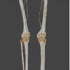
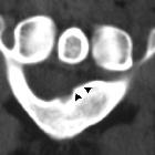
CT angiography can help identify abnormal tendinous or muscular structures, diastasis between the popliteal artery and vein, an insertion anomaly and/or arterial deviation. Aberrant muscular and fibrous attachments can be different in almost every case and it best evaluated on axial images.
MRI
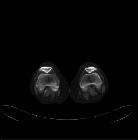
MRI is the best imaging modality to demonstrate the underlying anatomic type of entrapment, which helps guide surgical management . A medial slip of the medial head of the gastrocnemius may be seen, compressing the popliteal artery.
Angiography (DSA)
Lower limb angiography usually demonstrates medial deviation/compression of the popliteal artery when the ankle is plantarflexed. Occlusion of the vessels with thrombus can be seen in the acute presentation. Usually, collateral vessels are present. Even slight irregularity of the vessel can indicate a degree of entrapment .
Treatment and prognosis
Acute limb-threatening thrombosis requires urgent bypass surgery. Intermittent occlusion can usually be cured with the release of the popliteal artery alone or with saphenous vein bypass .
Siehe auch:
- Zystische Adventitiadegeneration
- Aneurysma der Arteria poplitea
- Dissektion
- Arteria poplitea
- vaskuläre Kompressionssyndrome
- Nervenkompressionssyndrom
- vaskuläre Syndrome
- thrombosiertes Aneurysma der Arteria poplitea
- Adduktorenkanalsyndrom
und weiter:
- Tibialis-anterior-Syndrom
- the value of MRI in popliteal artery entrapment syndrome
- carotid artery entrapment by hyoid bone
- popliteal artery entrapment syndrome with distal embolisation
- popliteal artery entrapment syndrome: multislice CTA vs DSA.
- popliteal artery entrapment by an aberrant muscular slip
- Gastrocnemius-Myalgie-Syndrom

 Assoziationen und Differentialdiagnosen zu Arteria-Poplitea-Kompressionssyndrom:


