Psoas hematoma


Psoas hematomas are located in the retrofascial space, rather than in the retroperitoneum, because the psoas muscles are located in the iliopsoas compartment posterior to the transversalis fascia, which is the posterior boundary of the retroperitoneum.
Clinical presentation
Presentation is often vague and non-specific and may include abdominal, pelvic, back or groin pain and/or swelling. There may be signs of hemorrhage including tachycardia, hypotension or fall in the measured hemoglobin level .
Psoas hematomas can present with constipation, urinary frequency, or fever if they are large. They can even compress the femoral nerve and cause a femoral neuropathy .
Pathology
There are many causes for psoas hematomas :
- spontaneous secondary to hemophilia, anticoagulation or atherosclerotic disease
- trauma
- ruptured abdominal aortic aneurysm (AAA); postoperative AAA repair; AAA endoleak
- post surgery or biopsy
- tumor
- inflammatory disease
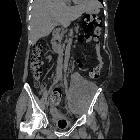
Radiographic features
Ultrasound
- acute hematoma - low echogenicity; may have a similar appearance to a cyst
- chronic hematoma - septa formation, calcification or appearance as a solid mass are all features
CT
- may present simply as an enlarged psoas muscle
- acute blood will present as an area of high density +/- fluid-fluid level
- chronic hematomas may have a similar appearance to psoas abscess
MRI
Appearance on MRI depends on the age of the hematoma :
- acute hematoma
- T1WI - isointense or slightly hypointense to muscle
- T2WI - may be hypointense or hyperintense
- subacute hematoma
- T1WI - high intensity rim, higher intensity peripheral zone and lower intensity core
- T2WI - relatively higher signal from core to periphery when compared to T1WI
- chronic hematoma
- hypointense rim on both T1WI and T2WI
Treatment and prognosis
Psoas hematoma can be complicated by superimposed infection or abscess and sometimes percutaneous aspiration and culture can be the only way to differentiate between the two .
Differential diagnosis
- psoas abscess
- necrotic mass
- retroperitoneal hemorrhage - lies anteriorly to the psoas muscle
Siehe auch:
- Psoasabszess
- retroperitoneales Hämatom
- Musculus psoas major
- spontane retroperitoneale Einblutung
- Marcumar
- Myositis ossificans im Musculus psoas
und weiter:


 Assoziationen und Differentialdiagnosen zu Hämatom im Musculus psoas: