Psoasabszess


Psoas muscle abscess and fluid collections are located in the retrofascial space, rather than in the retroperitoneal space, because the psoas muscles are located in the iliopsoas compartment posterior to the transversalis fascia, which is the posterior boundary of the retroperitoneum.
Clinical presentation
Psoas muscle abscess may present with fever, flank pain, abdominal pain or limp. Other symptoms are nausea, malaise and weight loss.
Pathology
Psoas muscle abscess may be classified as primary or secondary depending on the presence or absence of underlying disease.
Primary
Primary psoas muscle abscess occurs probably as a result of haematogenous spread of an infectious process from an occult source in the body. Primary psoas muscle abscess can occur in patients with:
- diabetes mellitus
- intravenous drug use
- AIDS
- renal failure
- immunosuppression
Secondary
Spread of infection from gastrointestinal disease (e.g. appendicitis, diverticulitis, Crohn disease or perforated colon carcinoma) is the most common source of a secondary psoas muscle fluid collection.
Renal disease is the second most common source.
They may also occur as a result of a neighboring spondylodiskitis.
Radiographic features
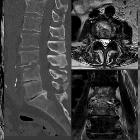
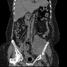
CT/MRI
Cross-sectional imaging is the modality of choice for abscess detection in the psoas muscle. Extension from the psoas muscle into the iliacus muscle is a common sequela.
Ultrasonography
It is diagnostic in only 60% of cases of psoas abscess, compared with 80% to 100% for CT.
Treatment and prognosis
Appropriate antibiotics along with drainage of the abscess are the treatment of choice. This is often treated with image-guided percutaneous drainage, typically CT, due to the retroperitoneal location.
Differential diagnosis
Siehe auch:
- Spondylodiscitis
- Spondylitis
- interventionelle Drainage Psoasabszess
- tuberkulöser Abszess Musculus iliopsoas
- Einblutung Musculus iliopsoas
- Hämatom im Musculus psoas
- AIDS
- psoas muscles
und weiter:


 Assoziationen und Differentialdiagnosen zu Psoasabszess: