Pulmonary cryptococcus infection


Pulmonary cryptococcosis is a form of pulmonary fungal infection caused by Cryptococcus gattii and C. neoformans. The respiratory tract is the principal route of entry for infection via inhalation of fungal spores.
For a general discussion of infection with this organism, please refer to the article cryptococcosis.
Epidemiology
Cryptococcosis predominantly occurs in immunocompromised patients but can also be seen in immunocompetent hosts, particularly, those exposed to avian (e.g. pigeon) droppings. The spectrum of pulmonary cryptococcosis depends on the host's defenses.
Clinical presentation
The presentation of pulmonary cryptococcosis can range from asymptomatic nodular disease to severe acute respiratory distress syndrome (ARDS) :
- most often, causes several lung nodules or masses +/- cavitation, chiefly in immunocompromised patients
- additionally, consolidation, mediastinal lymphadenopathy, and pleural effusion may also be present.
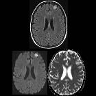
In the immunocompetent host the pulmonary infections normally are asymptomatic, in contradistinction to the immunocompromised patient, in whom cryptococcal infection is most often symptomatic, and commonly disseminates to the central nervous system, skin, and bones .
Overall, approximately one-third of patients are asymptomatic.
Symptoms range from a mild cough and low-grade fever to acute presentation with high fever and severe shortness of breath.
Pathology
The method of entry is usually by inhalation of cryptococcal particles into the lungs, causing pulmonary infection. Spores are found worldwide in soil contaminated by avian droppings.
Serology
Serum cryptococcal antigen (sCRAG) levels are helpful in diagnosis and follow-up.
Radiographic features
CT
In general, there are several CT patterns that can be seen:
- clustered nodular pattern: most prevalent
- solitary pulmonary nodule or mass with or without cavitation
- scattered nodules
- peribronchovascular consolidation
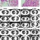
The most common CT findings in immunocompetent patients with pulmonary cryptococcosis are pulmonary nodules. The nodules are most often multiple, smaller than 10 mm in diameter, and well-defined with smooth margins. The nodules usually involve less than 10% of the parenchyma and tend to be distributed peripherally (up to 65% ) in the middle and upper zones. Where there are multiple nodules, they are usually bilateral . Associated cavitation may be seen in up to 40% of cases . Occasionally, unusual presentation such as large cavities may be seen .
Cavitations within nodules/masses tends to be more frequently present in immunocompromised patients than in immunocompetent patients .
Nuclear medicine
FDG PET-CT may play a complementary role to CT and ~60% of patients show higher FDG uptake than the mediastinal blood pool .
Treatment and prognosis
- antifungals such as oral fluconazole or intravenous amphotericin B
History and etymology
In 1924, Sheppe reported, for the first time, a case of pulmonary cryptococcosis .
Differential diagnosis
- pulmonary tuberculosis
- actinomycosis
- semi-invasive aspergillosis
- other pulmonary fungal infections
- primary lung neoplasia
- metastatic lung lesion
Siehe auch:
- acute respiratory distress syndrome (ARDS)
- Kavernöse Lungenläsionen
- Lungenrundherd
- Kryptokokkose
- pulmonale Pilzinfektionen
und weiter:


 Assoziationen und Differentialdiagnosen zu pulmonale Kryptokokkose: