pulmonary paragonimiasis
Pulmonary paragonimiasis is a food-borne parasitic disease caused by the lung fluke (trematode) Paragonimus westermani. It is endemic in southeast Asia, the Far East, and is also relatively common in Latin America and Africa . The disease can affect both the lung and pleura , although lung parenchymal involvement is thought to be commoner than pleural involvement .
Radiographic features
CT
CT chest: HRCT
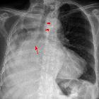
It usually manifests as a poorly marginated subpleural or subfissural nodule of about 2 cm in diameter that frequently contains a necrotic low attenuation area . The constellation of focal pleural thickening and subpleural linear opacities leading to a necrotic peripheral pulmonary nodule is another frequent CT finding.
There are also a myriad of other associated non-specific features. These can vary with the stage of disease.
Early findings are thought to be caused by the migration of juvenile worms and include:
- pneumothorax or hydropneumothorax
- focal airspace consolidation
- linear opacities
Later findings are thought to be caused by worm cysts and include:
- thin-walled cysts
- dense mass-like consolidation
- nodules
- bronchiectasis
Differential diagnosis
Imaging features can sometimes mimic that of lung cancer or pulmonary tuberculosis .
Siehe auch:


 Assoziationen und Differentialdiagnosen zu pulmonary paragonimiasis: