quadrigeminal cistern lipoma


Quadrigeminal cistern lipomas make up approximately 25% of intracranial lipomas and are located within the quadrigeminal cistern. They may be associated with hypoplasia of the inferior colliculus or agenesis of the corpus callosum.
For a general discussion please refer to the article on intracranial lipomas.
Clinical presentation
They are usually asymptomatic and are usually found incidentally. Rarely do they cause mass effect resulting in seizures or hydrocephalus .
Radiographic features
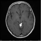
CT and MRI findings are characteristic for a fat-containing mass or lesion, however located in the quadrigeminal cistern.
CT
CT demonstrates a lobulated, non-enhancing fat density mass in the quadrigeminal cistern. Peripheral calcifications can be present in some cases.
MRI
MRI reveals signal characteristic of fat:
- T1: high signal intensity
- T2: high signal intensity
- T1 C+ (Gd): no enhancement
- fat saturated sequences: low signal intensity
- SWI: can produce blooming due to susceptibility artifact
Treatment and prognosis
Lipomas, in general, are mostly asymptomatic. If there is mass effect causing seizures or hydrocephalus, then surgical management can be considered .
Differential diagnosis
Differentials specific to its location (quadrigeminal palate) include tectal plate glioma or mass, tectal plate cyst, arachnoid cyst, dermoid cyst and epidermoid cyst .
See also
Siehe auch:
- Lipom
- intrakranielle Lipome
- Cisterna quadrigeminalis
- Tumoren der Pinealisregion
- Dysgenesie des Corpus callosum
- Tumoren des Tectums
und weiter:


 Assoziationen und Differentialdiagnosen zu tektales Lipom:

