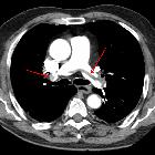
saddle pulmonary embolism

Computertomographie
mit intravenöser Kontrastmittelgabe mit Darstellung einer Embolie in den zentralen Lungenarterien (reitender Thrombus).

This image
is part of a series which can be scrolled interactively with the mousewheel or mouse dragging. This is done by using Template:Imagestack. The series is found in the category Pulmonary embolism - CT - case 001. Reitender Thrombus bei Lungenembolie. Genaugenommen sind es hier mehrere zentrale von links nach rechts reitende Thromben. Die linken Hauptstämme sind weitestgehend okkludiert.

A large
pulmonary embolism at the bifurcation of the pulmonary artery (saddle embolism).


CT pulmonary
angiogram (protocol) • Saddle pulmonary embolus - Ganzer Fall bei Radiopaedia
Saddle pulmonary embolism commonly refers to a large pulmonary embolism that straddles the bifurcation of the pulmonary trunk, extending into the left and right pulmonary arteries.
If large enough, it can completely obstruct both left and right pulmonary arteries resulting in right heart failure and, unless treatment is prompt, death.
With such extensive embolic burden, signs of right heart strain are usually present and include:
- dilatation of the right ventricle (RV) (i.e. RV width > left ventricular width)
- straightening or leftward bulging of the interventricular septum
- enlargement of the pulmonary trunk
Contrast reflux into the azygos vein, via the superior vena cava, and hepatic veins, via the inferior vena cava, is a controversial sign of RV strain, as it often occurs in the absence of raised right-sided heart pressures.
Siehe auch:

 Assoziationen und Differentialdiagnosen zu Reitender Thrombus:

