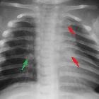
scapular fracture

Fraktur der
Scapula im Röntgenbild und in der Volumen-Rendering-Rekonstruktion der Computertomographie.


Scapular
fracture • Comminuted intra-articular glenoid fracture - Ganzer Fall bei Radiopaedia

Scapular
fracture • Bilateral humeral head avascular necrosis - Ganzer Fall bei Radiopaedia

Scapular
fracture • Scapular fracture - Ganzer Fall bei Radiopaedia

Scapular
fracture • Scapular fracture - Ganzer Fall bei Radiopaedia

Scapular
fracture • Acromial fracture - Ganzer Fall bei Radiopaedia

Scapular
fracture • Scapular and clavicle fractures - Ganzer Fall bei Radiopaedia

Scapular
fracture • Acromial fracture - ultrasound - Ganzer Fall bei Radiopaedia

Scapular
fracture • Scapular fracture (incidental) - Ganzer Fall bei Radiopaedia

Scapular
fracture • Glenoid rim fracture - Ganzer Fall bei Radiopaedia

Scapular
fracture • Scapular fracture - Ganzer Fall bei Radiopaedia

Scapular
fracture • Scapular fracture - Ganzer Fall bei Radiopaedia

Scapular
fracture • Scapular fracture - Ganzer Fall bei Radiopaedia
Scapula fractures are uncommon injuries, representing ~3% of all shoulder fractures.
Pathology
Mechanisms of injury
- requires high energy trauma (e.g. motor vehicle accidents account for 50% of scapular fractures)
- direct trauma to the shoulder region
- indirect trauma through falling on outstretched hand
- non-accidental injuries in children
Associations
Scapular fractures are often associated with other injuries:
- clavicle fracture
- rib fracture
- sternal fracture
- spinal fracture
- pneumothorax and/or pulmonary contusion
- brachial plexus injury
Radiographic features
Plain radiograph
Requires trauma series views to demonstrate the fractures due to the superimposition of the shoulder girdle and thoracic cage.
Radiographic series includes:
CT
- standard for diagnosis and evaluation of the fracture and its associated injuries
- axial scan with coronal, sagittal and 3D reconstructions are used in the assessment of scapular injuries
Classification
- intra-articular glenoid fracture
- type I: avulsion of anterior glenoid margin
- type II: transverse or oblique fracture through glenoid fossa exiting inferiorly
- type III: oblique fracture through glenoid fossa exiting superiorly and associated with acromioclavicular joint injury
- type IV: transverse fracture exiting through the medial scapular border
- type V: combination of type II and type IV
- type VI: comminuted glenoid fracture
- extra-articular glenoid fracture
- type I: glenoid neck fracture without clavicular fracture
- type II: glenoid neck fracture with clavicular fracture and acromioclavicular dislocation
- coracoid process fracture
- type I: fracture proximal to the coracoclavicular ligament
- type II: fracture distal to the coracoclavicular ligament
- acromial fracture
- type I: minimally displaced
- type II: displaced but does not reduce subacromial space
- type III: displaced and narrow the subacromial space
Differential diagnosis
- os acromiale and other accessory ossicles of the scapula
Siehe auch:
und weiter:

 Assoziationen und Differentialdiagnosen zu Scapulafraktur:

