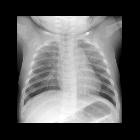
rib fracture


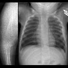


Rib fractures are a common consequence of trauma and can cause life-threatening complications.
Epidemiology
Associations
Rib fractures are often associated with other injuries and the greater the number of rib fractures the more likely are associated injuries :
- brachial plexus or subclavian vessel injuries (1-3 rib fractures
- pneumothorax/hemothorax
- pulmonary laceration
- lung herniation
- liver, kidney and spleen traumatic injuries (10-12 rib fractures)
Pathology
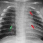
The 4-10 ribs are the most commonly fractured . Fractures of the 1-3 ribs are associated with high-energy trauma .
When the rib is fractured twice, the term floating rib is used to describe the free fracture fragment, and when three or more contiguous floating ribs are present this is called a flail chest.
Buckle rib fractures can also occur.
Etiology
- blunt and penetrating trauma: e.g. motor vehicle accidents, falls, assaults
- most common injury in blunt thoracic trauma, occurring in 50% of cases
- pathological fractures
- stress fractures: occur more commonly in high-level athletes
- non-accidental injuries in children
- typically posterior fractures
- intimate partner violence
- typically posterior
- cardiopulmonary resuscitation (CPR): rib fracture occurs in 1 in 3
- fetal rib fractures: caused by skeletal dysplasias
- radiation-induced rib fractures
- spontaneous: spontaneous rib fracture
Radiographic features
Plain radiograph
- may miss up to 50% of rib fractures even with dedicated oblique rib projections
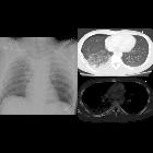
CT
- more sensitive than plain radiography for the detection of rib fractures
- Displacement on CT may be defined as greater than half a rib shaft width
Ultrasound
Common application of point of care ultrasonography, used in a complementary manner to conventional radiography in the workup of blunt chest wall trauma and localized chest pain. Ultrasonography is more sensitive and specific than conventional radiography for rib fracture detection in blunt trauma when performed by a trained clinician .
Moreover, the use of sonography allows the detection of radiographically occult costal cartilage fractures, as well as assessment of the underlying lung for associated traumatic pathology, including;
B-mode
- cardinal sonographic feature is a discontinuity of the otherwise smooth, linear echogenicity corresponding to the anterior bony cortex
- if accompanied by cortical displacement, may be further characterized by the axial distance between the cortical margins
- separation by less than 1 mm defines mild displacement
- considered severe when >4 mm
- often associated with comet tail artifacts extending from the fracture site into the far field
- if initially occult, obvious interruption of the bony cortex may be elicited with sonopalpation (applying local transducer pressure)
- appropriate analgesia may be necessary before attempting this maneuver
- suggestive findings include a circumscribed, hypoechoic fluid collection adjacent to the putative fracture site
- represents a fracture hematoma, fluid nature may be ascertained through careful adjustment of gain settings and dynamic variation of the observed shape with pressure
- associated periosteal elevation appears as a delicate, raised hyperechoic linear stripe
Nuclear medicine
- Tc-99m bone scan is sensitive but not specific for rib fractures and demonstrates focal areas of high-uptake, which need to be correlated with SPECT or radiographic imaging
Treatment and prognosis
Rib fractures themselves are treated symptomatically and have a good prognostic outcome. Rarely, severe rib injuries (e.g. flail chest) may be treated with ORIF, often in the setting of other severe traumatic injuries and in the hope that respiratory function will improve facilitating a shorter ICU stay and quicker recovery.
Complications
Aside from immediate traumatic complications outlined above atelectasis and pneumonia may develop, mainly due to poor respiratory effort secondary to pain, and this increases the morbidity and mortality due to rib fractures .
See also
Siehe auch:
- flail chest
- Läsionen der Rippen
- buckle rib fracture
- Fraktur Rippenknorpel
- interkostale Lungenhernie
- Rippenklammern
- kindliche Rippenfrakturen
- Rippenserienfraktur
- Rippenfraktur Sonographie
- Rippenfrakturen in der Szintigraphie
- Brustwandrekonstruktion
- Rippenfraktur Verlaufskontrolle
- isolated 1st rib fracture in a child
- Rippenpseudarthrose
- fetale Rippenfrakturen
und weiter:


 Assoziationen und Differentialdiagnosen zu Rippenfraktur: