Sinusitis ethmoidalis

A rare case
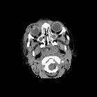
of sinolith in the ethmoid sinus. Axial and coronal CT images reveal a calcified, hyperdense irregularly-shaped mass in the left ethmoid region bulging into the sphenoid sinus. Inflammatory tissue in bilateral ethmoidal air cells was also noted.

A rare case
of sinolith in the ethmoid sinus. Axial and coronal CT images reveal a calcified, hyperdense irregularly-shaped mass in the left ethmoid region bulging into the sphenoid sinus. Inflammatory tissue in ethmoidal air cells was also noted.

A rare case
of sinolith in the ethmoid sinus. MRI shows a signal void in the left ethmoid region on both T1 and T2 weighted axial images. Also inflammatory tissue in ethmoid air calls is demonstrated.

A rare case
of sinolith in the ethmoid sinus. MRI shows a signal void in the left ethmoid sinus on both T1 and T2 weighted axial images. Also inflammatory tissue in ethmoidal air cells is demonstrated.

School ager
with right eyelid swelling and redness and right eye pain. Axial CT with contrast of the orbits (above) shows opacification of the right ethmoid sinus, inflammatory changes within the right orbit, and a low density oval fluid collection with an enhancing wall between the right medial rectus muscle and the right ethmoid sinus. Coronal CT (below) shows how this fluid collection is displacing the right medial rectus muscle laterally.The diagnosis was right ethmoid sinusitis causing right orbital cellulitis and subperiosteal abscess.
Sinusitis ethmoidalis
Sinusitis Radiopaedia • CC-by-nc-sa 3.0 • de
Sinusitis is a broad and non-specific term referring to the inflammation within the paranasal sinuses. There are several forms which are specific entities based on etiology and clinical features, and hence covered individually:
Siehe auch:


 Assoziationen und Differentialdiagnosen zu Sinusitis ethmoidalis: