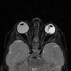
Sonographie des Auges

Ocular
ultrasonography focused on the posterior eye segment: what radiologists should know. Optic disc drusen. a Axial US shows a typical calcified plaque at the optic disc (arrow). b Drusen typically exhibit autofluorescence

Ocular
ultrasonography focused on the posterior eye segment: what radiologists should know. Diagram illustrating ocular anatomy. The anterior segment comprises the cornea (1), anterior chamber (AC), iris (2), ciliary body (3), lens, and posterior chamber (PC). The AC and PC are filled with aqueous humour. The lens is laterally attached to the ciliary body. The posterior segment comprises the vitreous chamber (4) and the posterior ocular wall (5), which is formed by the retina, choroid, and sclera (posterior RCS complex). The vitreous chamber is filled with vitreous humour, and its periphery is called the vitreous capsule or hyaloid. The retina anchors anteriorly at the ora serrata (curved arrow) and posteriorly at the optic disc (6). The choroid anchors anteriorly at the scleral spurs near the ciliary bodies and posteriorly near the exit foramina of the vortex veins (at some distance anterior to the optic disc). Behind the globe, the optic nerve (ON) is seen.

Ocular
ultrasonography focused on the posterior eye segment: what radiologists should know. Posterior vitreous detachment (PVD) in two patients. a, b Illustration and US image correlation showing a PVD. Note the undulating appearance of the posterior vitreous that is better identified on dynamic scanning (arrowheads). c Axial US in a 67-year-old man with blurred vision. Note the fine echoes in the retrohyaloid space (asterisk), consistent with retrohyaloid haemorrhage

Ocular
ultrasonography focused on the posterior eye segment: what radiologists should know. Choroidal hemangioma in a 45-year-old woman with choroidal hemangioma suspected at fundoscopy. Axial US shows a small homogeneous hyperechoic biconvex lesion (arrow) on the temporal side of the globe near the papilla

Ocular
ultrasonography focused on the posterior eye segment: what radiologists should know. Neovascular age-related macular degeneration. a Top image: optical coherence tomography (OCT) image in a patient with active neovascular age-related macular degeneration (AMD) shows serous detachment of retinal pigment epithelium (arrows) and neurosensory retinal detachment (arrowheads). Note also the subretinal haemorrhage (white circle) and subretinal fluid (white asterisk). Bottom image: non-pathologic OCT in another patient. b 71-year-old man with progressive vision loss in the right eye. US shows a mass on the temporal side of the posterior wall next to the papilla (asterisk); note also the posterior vitreous detachment (arrows). Final diagnosis was subretinal haemorrhage due to choroidal neovessels in the context of neovascular AMD

Ocular
ultrasonography focused on the posterior eye segment: what radiologists should know. Choroidal melanoma in two patients. a 62-year-old man with a 6-month history of progressive vision loss in the right eye. Fundoscopy image shows a pigmented mass in the inferior nasal arcade of the right eye (asterisk). b Colour Doppler US image shows a dome-shaped hypoechoic posterior mass with a smooth surface and choroidal excavation underneath (arrow), as well as internal blood flow. c, d US obtained in a 54-year-old man shows a mushroom-shaped mass related to a rupture of Bruch’s membrane. Note the peritumoral serous retinal detachment (arrowheads) induced by the growth of the tumour, as illustrated in the diagram

Ocular
ultrasonography focused on the posterior eye segment: what radiologists should know. Choroidal detachment. a Diagram showing typical bilateral exudative choroidal detachment, with two convex rigid lines protruding into the vitreous, extending from the scleral spur near the ciliary body (arrows) to the level of the exit foramina of the vortex veins (arrowheads), at some distance from the papilla. b US image in a 69-year-old woman with glaucoma in the right eye 1 day after cataract surgery shows two lines ballooning into the vitreous, demonstrating an exudative choroidal detachment (arrows)

Ocular
ultrasonography focused on the posterior eye segment: what radiologists should know. Proliferative diabetic retinopathy (PDR). a Fundoscopy image shows extensive neovascularization (arrows), the hallmark of PDR. b US images in a 72-year-old diabetic man with PDR and a history of recurrent vitreous haemorrhage. Note the vitreous bands due to chronic vitreous haemorrhage (arrows) and preretinal fibrovascular membranes (arrowhead). c At the 3-month follow-up, the fibrovascular tissue has grown into the vitreous cavity (arrowhead), and the patient has developed posterior vitreous detachment (arrow), retrohyaloid haemorrhage (asterisk), and focal retinal detachment (curved arrow). He subsequently underwent vitrectomy and panretinal laser photocoagulation

Ocular
ultrasonography focused on the posterior eye segment: what radiologists should know. Woman with chronic right retinal detachment who developed a retinal cyst (arrow) on the nasal side

Ocular
ultrasonography focused on the posterior eye segment: what radiologists should know. An 83-year-old woman with uncontrolled diabetes and a history of proliferative diabetic retinopathy and vision loss in the left eye. Axial US image shows thick membranes (arrowheads) in the vitreous and a tractional total chronic retinal detachment (arrows)

Ocular
ultrasonography focused on the posterior eye segment: what radiologists should know. Subretinal haemorrhage. Axial US shows the subretinal space (asterisks) filled with an echogenic material consistent with blood

Ocular
ultrasonography focused on the posterior eye segment: what radiologists should know. Retinal detachment. a Diagram illustrating total retinal detachment. Note the anatomic retinal attachment points. b Axial US image in a 45-year-old man with Marfan syndrome: two echogenic lines form an acute angle, with the “V” shape characteristic of an acute retinal detachment. At this stage, the retinal leaves are thin and mobile (arrows). This condition requires surgical treatment. c 75-year-old woman with chronic retinal detachment. At this stage, the retinal leaves look thick and rigid, adopting a funnel-shaped configuration (arrowheads); this condition is not amenable to surgical treatment

Ocular
ultrasonography focused on the posterior eye segment: what radiologists should know. Acute and chronic vitreous haemorrhage in two different patients. a 72-year-old man with history of diabetes mellitus who presented with sudden blurred vision due to acute vitreous haemorrhage. At US, subtle echogenic material that obscured the ophthalmoscopic examination is seen floating within the vitreous. b Axial US image in a 58-year-old man with a history of vitreous haemorrhage 4 months earlier shows echogenic bands in the vitreous representing fibrous membranes, characteristic of chronic vitreous haemorrhage. The patient went on to undergo vitrectomy

Ocular
ultrasonography focused on the posterior eye segment: what radiologists should know. Sonographic appearance of the structures of the normal eye. The cornea (1) is visualized as the most superficial echogenic curved line; the anterior chamber (2) is anechoic. The iris (3) appears as a thin echogenic line. The lens (4) is defined by anterior and posterior boundary echoes, but the lens itself is echo-free. The vitreous chamber (5) is filled with a clear gel-like substance that is normally echo-free, although the formation of spots and linear echoes with aging is considered normal. The RCS complex (6) forms the wall of the posterior ocular segment; it is seen as an echogenic concave line extending from the iris plane to the optic nerve (ON; 7). The ON is seen as a hypoechoic band surrounded by echogenic retrobulbar fat (8). The circular area where the ON connects to the retina is the optic disc or papilla (9)

Ocular
ultrasonography focused on the posterior eye segment: what radiologists should know. Asteroid hyalosis. Axial US in a 58 year–old man without visual disturbances. Note the incidental presence within the vitreous of numerous small hyperechoic echoes with comet-tail artefacts

Ocular
ultrasonography focused on the posterior eye segment: what radiologists should know. Ocular US in a 25-year-old woman with trisomy 21 and persistent hyperplastic primary vitreous. Axial US image shows a right eye that is smaller than normal, with a linear echogenic tract extending from the posterior surface of the lens to the posterior ocular wall representing the fibrovascular remnant (arrow). Colour Doppler shows arterial blood flow within the band that corresponds to persistence of the hyaloid artery (dotted arrow). Note also the cataract

Ocular
ultrasonography focused on the posterior eye segment: what radiologists should know. Complete lens dislocation in a man with a history of ocular trauma. Axial US image shows the dislocated lens (arrow), which lies entirely within the vitreous. Membranes from associated vitreous haemorrhage can also be seen

Ocular
ultrasonography focused on the posterior eye segment: what radiologists should know. Partial lens dislocation in a 45-year-old man with a history of ocular trauma 2 years earlier. He developed a post-traumatic cataract, and physical examination detected an abnormal vibration or agitated motion of the iris during eye movements—the so-called iridodonesis (movie 1). a US examination with the patient in a supine position showed a globulous lens situated in a normal position, as well as cataracts. b US examination with the patient sitting upright revealed that the lens was detached at the 12 o’clock position. It is sometimes useful to perform US studies with the patient in different positions

Ocular
ultrasonography focused on the posterior eye segment: what radiologists should know. A 74-year-old man with a history of ocular trauma 20 years earlier. Axial US image of the right eye shows increased echogenicity of both walls of the lens, with intraocular echoes. There is also a synechia between the iris and the lens, causing the iris to curve forward (arrows)—the so-called iris bombe. This condition prevents the flow of aqueous humour from the posterior to the anterior chamber. Also note the thick posterior membranes between the lens and the vitreous (arrowheads)

Ocular
ultrasonography focused on the posterior eye segment: what radiologists should know. Unilateral cataracts in a 73-year-old man. Compare the intralenticular echoes in the right eye (a) with the normal appearance of the lens in the left eye (b)

Ocular
ultrasonography focused on the posterior eye segment: what radiologists should know. Phthisis bulbi in a 63-year-old man with a history of left ocular trauma during infancy. Axial US image shows a small, crenated, shrunken-looking ocular globe with calcified walls (arrows). Cataracts and partial lens dislocation (asterisk) are also seen

Ocular
ultrasonography focused on the posterior eye segment: what radiologists should know. Comparative study in a woman with anisometropia in long-standing myopia in the right eye. a. Longitudinal US shows the lengthened anteroposterior axis of the right eye, with a pear-shaped posterior pole sacculation known as a staphyloma (arrow). b Compare with the normal left globe

Ocular
ultrasonography focused on the posterior eye segment: what radiologists should know. US technique. Photograph from a standard examination shows the position of the transducer to acquire axial images of the eye. Gel should be applied abundantly to allow better contact between the transducer and the eye

Ocular
ultrasonography focused on the posterior eye segment: what radiologists should know. Normal fundoscopy of the right eye shows the oval optic disc (1); retinal veins (2) and arteries (3) radiate from the centre. The macula (4) is the round dark area lateral to the disc on the temporal side of the eye. The macula has a depressed spot, the fovea centralis (asterisk), which is the area of most acute vision

Ocular
ultrasonography focused on the posterior eye segment: what radiologists should know. Post-surgical conditions. a Patient with pseudophakia with an intraocular lens. Note the reverberation artefact produced by the intraocular lens (arrows). b US appearance of scleral buckling (arrows) used to treat retinal detachment (RD). Note the absence of the lens in this aphakic patient (arrowhead). c US showing silicone oil injection and scleral buckling, with artefacts produced by the silicone. d Reverberation artefact caused by retention of a small volume of perfluorocarbon liquid in the vitreous cavity (arrows) in a man treated for RD
Sonographie des Auges
Siehe auch:
- Aderhautmelanom
- Staphylom
- Drusen der Papilla nervi optici
- Linsenluxation
- Netzhautablösung
- Glaskörperblutung
- persistierender hyperplastischer primärer Glaskörper (PHPV)
- Aderhautablösung
- Aderhauthämangiom
- Katarakt
- Phthisis bulbi oculi
- posterior lenticonus
- Glaskörperabhebung
- Makuladegeneration
- Verletzungen Bulbus
- künstliche Augenlinse
- Asteroide Hyalose
- subretinale Einblutung
- retinale Zyste
- proliferative diabetische Retinopathie

 Assoziationen und Differentialdiagnosen zu Sonographie des Auges:
persistierender
hyperplastischer primärer Glaskörper (PHPV)


