persistierender hyperplastischer primärer Glaskörper (PHPV)


Persistent hyperplastic primary vitreous (PHPV), also known as the persistent fetal vasculature, refers to a rare congenital developmental malformation of the eye.
Clinical presentation
Clinically, this condition usually manifests as unilateral or bilateral leucocoria. Patients may also have poor vision, small eye (microphthalmia) and strabismus.
Pathology
It arises due to a failure of normal regression of the embryonic hyaloid vascular system. In the normal situation the primary vitreous forms around the 7 week of gestation life and starts involuting around 20 week and nearly always disappears at the time of birth. Persistent fetal vasculature in PHPV can lead to fibrosis, resulting in elongation of the ciliary processes, retinal detachment, and spontaneous cataracts.
Subtypes
PHPV can be divided into either anterior (ventral) or posterior (dorsal) types, with most patients with PHPV having a combination of these .
Associations
PHPV can occur on its own or association with various other conditions notably when bilateral, these include :
- Norrie disease
- Warburg syndrome
- retinal dysplasia
Radiographic features
From an imaging standpoint, only the features of posterior PHPV are well known .
General
In posterior PHPV, the globe is small and can contain retinal detachments.
Ocular ultrasound
An echogenic band may be seen in the posterior segment of the globe extending from the posterior surface of the lens to the optic nerve head. On color Doppler, arterial blood flow may be seen within this band.
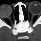
CT
The CT appearance can be quite variable and the described spectrum of CT findings includes :
- soft-tissue replacement (infiltration) of the vitreous body
- retrolental soft tissue along the Cloquet canal: fine linear structure
extending from the head of the optic nerve to the posterior surface of the lens - absence of abnormal calcification within the orbit
- microphthalmus
- retrohyaloid layered blood
- hypervascularity of the vitreous humor: after contrast administration, the vitreal abnormalities may enhance, which is believed to reflect a persistent hypervascular vitreous
- retinal detachments can be hyperdense on CT
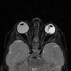
MRI
On MRI the retrolenticular tissue characteristic of this condition has a triangular shape, like that of a martini glass appearing as low T2 signal against the normal high T2 signal of the globe .
Differential diagnosis
- clinically it is one of the more significant and frequent conditions that can mimic a retinoblastoma
- at ultrasound the primary differential is of retinal detachment
Siehe auch:
- Pätau-Syndrom
- Retinoblastom
- Netzhautablösung
- Leukokorie
- Walker-Warburg-Syndrom
- Glaskörperblutung
- Retinale Dysplasie
- Glaskörperfehlbildungen
- Norrie-Syndrom
- microphthalmus
und weiter:


 Assoziationen und Differentialdiagnosen zu persistierender hyperplastischer primärer Glaskörper (PHPV):