Warburg syndrome

Prenatal
presentation of Walker–Warburg syndrome with a POMT2 mutation: an extended fetal phenotype. Fetus at 20 weeks’ gestational age with ultrasound findings of Walker Warburg syndrome. a Three-dimensional surface rendering of the fetal face showing bilateral microphthalmia and a clenched hand. b Axial US through the orbits showing asymmetric affection with the left eye demonstrating homogeneous opacity of the lens in the left eye consistent with cataract, and the right eye showing echogenic band consistent with hyperplasia of the vitreous chamber. c Axial trans-ventricular plane of the fetal head demonstrating agenesis of corpus callosum, dilatation of the third ventricle, and the anterior and posterior horns of the lateral ventricles. d Axial transcerebellar plane of the fetal head showing cerebellar hypoplasia and vermian agenesis with open fourth ventricle communicating with the cisterna magna and non-expanded posterior fossa. e Fetal echocardiogram demonstrating AVSD (atrial ventricular septal defect). f Coronal US image of multicystic dysplastic kidney (MCDK) showing enlarged kidney with several subcortical small cysts. g Three-dimensional surface rendering mode of lower extremities showing hyperextended knees and bilateral talipus with deformed lower limbs. h Postmortem image confirms the presence of the above findings
Walker-Warburg syndrome (WWS), sometimes known as HARDE syndrome, is an extremely rare lethal form of congenital muscular dystrophy. It is primarily characterized by:
- fetal hydrocephalus: almost always present
- neuronal migrational anomalies: agyria (cobblestone lissencephaly / lissencephaly type II)
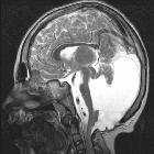
- distinctive dorsal "kink" at the mesencephalic-pontine junction
- Dandy Walker continuum
- retinal dysplasia
- encephalocoele
- cerebellar malformations
- congenital muscular dystrophy
Additional anomalies include:
- agenesis of the corpus callosum
- microphthalmia or unilateral buphthalmos
- ocular colobomas
- microtia
- congenital cataracts
- genital anomalies in males
- cleft lip +/- palate
Pathology
Genetics
It is considered by many to be inherited in an autosomal recessive manner.
Radiographic features
MRI
Different brain abnormalities can be present, such as:
- diffuse cobblestone cortex
- complete absence of cerebral and cerebellar myelin
- cerebellar polymicrogyria
- pontine and cerebellar vermal hypoplasia
- hydrocephalus
- variable callosal hypogenesis
Treatment and prognosis
The overall prognosis is poor with most infants dying within the 1year of life. There is no specific treatment and management is mainly supportive.
History and etymology
It was named after Arthur Earl Walker and Mette Warburg.
Siehe auch:
- Lissenzephalie
- Lippen-Kiefer-Gaumen-Spalte
- Meningoenzephalozele
- Kolobom
- Mikrophthalmus
- Dysgenesie des Corpus callosum
- Lissenzephalie Typ 2
- Dandy Walker continuum
- Ohrmuschelfehlbildung
- Dystroglykanopathie
- kongenitaler Hydrozephalus
- Kongenitale Muskeldystrophie-Dystroglykanopathie mit Fehlbildungen des Gehirns und der Augen Typ A3
- Kongenitale Muskeldystrophie-Dystroglykanopathie mit Fehlbildungen des Gehirns und der Augen Typ A6
- Kongenitale Muskeldystrophie-Dystroglykanopathie mit Fehlbildungen des Gehirns und der Augen Typ A4
- Kongenitale Muskeldystrophie-Dystroglykanopathie mit Fehlbildungen des Gehirns und der Augen Typ A2
- Kongenitale Muskeldystrophie-Dystroglykanopathie mit Fehlbildungen des Gehirns und der Augen Typ A1
- Kongenitale Muskeldystrophie-Dystroglykanopathie mit Fehlbildungen des Gehirns und der Augen Typ A5
und weiter:

 Assoziationen und Differentialdiagnosen zu Walker-Warburg-Syndrom:


