Sprunggelenk


The ankle joint (also known as the tibiotalar joint or talocrural joint) forms the articulation between the foot and the leg. It is a primary hinge synovial joint lined with hyaline cartilage.
Gross anatomy
The ankle joint is comprised of the tibia, fibula and talus as well as the supporting ligaments, muscles and neurovascular bundles. It carries the weight of the body and can undergo a myriad of pathology, most commonly traumatic injuries of the medial and lateral malleoli.
The tibia extends inferiorly to articulate with the talus on its medial aspect which has an inferior projection at its medial aspect, the medial malleolus.
The fibula has a similar inferior projection laterally, the lateral malleolus. The tibia has a partially curved surface to articulate with the talar dome which is wide anteriorly and narrows posteriorly.
The talus lies superior to the calcaneus and articulates with the navicular anteriorly.
Ligaments
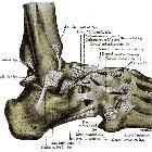
The ligaments of the ankle form a medial and lateral group each comprising of three main ligaments.
The deltoid ligament is medial and is made of two parts. The deep parts is the tibiotalar part. The superficial triangular (delta) part is a continuous band projecting from the apex of the medial malleolus to the medial tubercle of the talus, the sustentaculum tali of the calcaneus and the tuberosity of the navicular. With fusion with the spring ligament also.
The lateral ligaments include the anterior and posterior talofibular ligament and the calcaneofibular ligament. These ligaments fuse with the joint capsule to enclose the joint so any fracture involving the joint will invoke an ankle effusion. The interosseous tibiofibular ligament between the fibula and tibia is another important structure maintaining ankle stability. In the subtalar region the spring ligament (calcaneonavicular) maintains the integrity of the region.
Compartments
There is an anterolateral, posteromedial and lateral compartment of the ankle typically superficial to the joint.
The posteromedial compartment, in order of anterior to posterior has the tendons of tibialis posterior and flexor digitorum longus, the posterior tibial artery, the tibial nerve and flexor hallucis longus tendon.
The anterior compartment under the extensor retinaculum is the tibialis anterior tendon, extensor hallucis longus tendon, dorsalis pedis artery, deep peroneal nerve, extensor digitorum longus tendon.
In the peroneal compartment consists of peroneus longus, peroneus brevis, the sural nerve and the terminal branch of peroneal artery. The anteromedial superficial area is the long saphenous vein and saphenous nerve. Superficial to the peroneal compartment is the sural nerve and small saphenous vein.
Nerve supply
Blood supply
Siehe auch:
- Bänder des Sprunggelenkes
- Arthrose oberes Sprunggelenk
- Oberes Sprunggelenk
- Röntgen Projektion nach Saltzman
- Unteres Sprunggelenk Anatomie
- Distale tibiofibulare Syndesmose
- Sprunggelenk-Endoprothese
- Sehnenfächer Sprunggelenk
und weiter:

 Assoziationen und Differentialdiagnosen zu Sprunggelenk:


