Water-lily sign (hydatid cyst)

Primary renal
hydatid cyst. A thick-walled cystic lesion (red arrow) can be seen arising from interpolar region of right kidney (yellow arrow). The lesion shows a few internal non-enhancing membranes (green arrow) and daughter cysts (elbow arrows).

Water-lily
sign (hydatid cyst) • Hepatic hydatid cysts - Ganzer Fall bei Radiopaedia

Detachment of
the endocyst combined with biliary communicating rupture in hydatid disease. T2 weighted image with fat suppression showing a wall defect (arrow) in the anterior aspect of the cystic mass. The low-signal intensity rim is interrupted at this point.

Rare case of
echinococcosis with negative histology and cytology. The left hemithorax in coronal plane. There is an impression of the waterlily-sign in the fluid of the cyst.

Water-lily
sign (hydatid cyst) • Hepatic hydatid cyst - water lily sign - Ganzer Fall bei Radiopaedia

Water-lily
sign (hydatid cyst) • Peritoneal hydatid cyst mimicking a neoplasm - Ganzer Fall bei Radiopaedia

Water-lily
sign (hydatid cyst) • Hepatic hydatid cyst - Ganzer Fall bei Radiopaedia

Water-lily
sign (hydatid cyst) • Rupture of massive pulmonary hydatid cyst with tension hydropneumothorax - Ganzer Fall bei Radiopaedia

Water-lily
sign (hydatid cyst) • Calcified hepatic hydatid cyst - Ganzer Fall bei Radiopaedia

Water-lily
sign (hydatid cyst) • Hepatic hydatid cyst - ultrasound water lily sign - Ganzer Fall bei Radiopaedia

Primary renal
hydatid cyst. A thick-walled cystic lesion (red arrow) can be seen. It indents the liver parenchyma. The lesion shows a few internal non-enhancing membranes (green arrow) as well as daughter cysts (elbow arrows).

Water-lily
sign (hydatid cyst) • Hepatic hydatid cyst - Ganzer Fall bei Radiopaedia

Water-lily
sign (hydatid cyst) • Pulmonary hydatid cyst - Ganzer Fall bei Radiopaedia

Water-lily
sign (hydatid cyst) • Pulmonary hydatid cyst - Ganzer Fall bei Radiopaedia

Renal cystic
echinococcosis; an unusual localization of echinococcosis. Sagittal CT image of the right kidney,shows the lesion in the lower pole of the kidney.

Renal cystic
echinococcosis; an unusual localization of echinococcosis. Contrast-enhanced CT axial image; shows mild rim enhancement in the lesion.

Renal cystic
echinococcosis; an unusual localization of echinococcosis. Non-enhanced axial CT image; shows a low attenuating mass with multiple internal floating membranes.

Primary renal
hydatid cyst. A thick-walled cystic lesion(red arrow) can be seen. The lesion shows a few internal membranes (green arrow) as well as daughter cysts (elbow arrow).

Detachment of
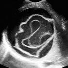
the endocyst combined with biliary communicating rupture in hydatid disease. US image showing a fluid collection with "floating membranes" representing the detached endocyst.
 nicht verwechseln mit: Linguini-Zeichen
The water-lily sign, also known as the camalote sign, is seen in hydatid infections when there is detachment of the endocyst membrane which results in floating membranes within the pericyst that mimic the appearance of a water lily.
It is classically described on plain radiographs (mainly chest X-ray) when the collapsed membranes are calcified but may be seen on ultrasound, CT, and MRI.
Siehe auch:
und weiter:

 Assoziationen und Differentialdiagnosen zu Wasserlilien-Zeichen:

