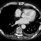
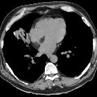
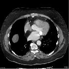
lipomatous hypertrophy of the interatrial septum


Lipomatous hypertrophy of the interatrial septum (LHIS) is a relatively uncommon disorder of the heart characterized by benign fatty infiltration of the interatrial septum. It is commonly found in elderly and obese patients as an asymptomatic incidentally discovered finding.
Epidemiology
The prevalence of LHIS is estimated to be between 1-8%. The incidence increases with age, body mass and chronic corticosteroid therapy. There may be a higher incidence in women .
Clinical presentation
The condition is usually asymptomatic and incidentally discovered in a majority of individuals . There may be arrhythmias or superior vena cava syndrome if the superior vena cava is encased.
Pathology
Microscopically, lipomatous hypertrophy of the interatrial septum (LHIS) is characterized by fat infiltration between the myocardial fibers of the atrial septum. LHIS also can create a masslike bulge. There is typical sparing of the fossa ovalis , with LHIS lying anterior to the fossa ovalis. It does not have a capsule, in contrast to cardiac lipoma.
Associations
- diffuse mediastinal lipomatosis
- increased epicardial fat
- long-term prednisone therapy in at least one case
Radiographic features
CT
Diagnosis is made with CT when a smooth, non-enhancing, well-marginated expansion of the interatrial septum is identified exceeding 2 cm in transverse diameter.
Because it spares the fossa ovalis, the lesion characteristically takes on a dumbbell-shaped appearance. This may best be seen on reconstructed short axis views.
Cardiac MRI
MRI diagnosis is straightforward in classic cases, and this entity is characterized by a bilobar interatrial septal thickening revealing homogeneous high signal intensity similar to that of subcutaneous fat tissue.
- T1: high signal
- T2 non-fat suppressed: high signal
- T2 fat suppressed: low signal
The exclusively fatty nature of such masses can be seen on fat-suppressed imaging.
FDG PET-CT
Usually shows a moderate degree of FDG-uptake. It may vary on serial studies and the FDG-avidity was initially thought to be caused by the metabolic activity of brown adipose tissue (BAT) . More recently this theory has been questioned by other authors, with suggestion of either inflammation or a hitherto unidentified mechanism of BAT activation as potential mechanism .
Be that as it may, there is consensus on the necessity of hybrid imaging and correlation with former imaging studies to avoid misclassification.
Treatment and prognosis
It is benign in many individuals and often does not warrant any treatment, in situations where there is severe superior vena cava obstruction or intractable rhythm disturbance, surgical excision with reconstruction of the interatrial septum is sometimes considered.
Although histologically benign, LHIS has been associated with adverse clinical sequelae including
- cardiac rhythm disturbances : lipomatous hypertrophy is usually situated in the area of at least two described interatrial conduction pathways (anterior and middle internodal pathways), and their interruption could be the major reason for rhythm disorders such as
- supraventricular arrhythmias
- atrial fibrillation
- atrial premature contractions
- atrioventricular block
- obstructive flow symptoms including dyspnea - especially if large
- syncope
- sudden death
History and etymology
It was first described by the American pathologist John T Prior (1917-2007) working at Syracuse College of Medicine, New York City, in 1964 .
Differential diagnosis
Differential diagnoses should be thought of when there is sparing of the fossa ovalis.
Fat-containing neoplasms that can arise in the atrial septum include:
See also
- fat containing thoracic lesions
- cardiac lipomatoses
Siehe auch:
- Vorhofmyxom
- Tumoren des Herzens
- Rhabdomyom des Herzens
- kardiales Lipom
- kardiales Liposarkom
- Rhabdomyosarkom des Herzens
und weiter:

 Assoziationen und Differentialdiagnosen zu Lipomatöse Hypertrophie des interatrialen Septums:


