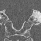
Osteom des Mittelohrs


Osteom des Mittelohrs
Osteom Radiopaedia • CC-by-nc-sa 3.0 • de
Osteomas are benign mature bony growths, seen almost exclusively in bones formed in membrane (e.g. skull).
Terminology
When they arise from bone they may be referred to as a "homoplastic osteoma", and when they arise in soft tissue they may be referred to as a "heteroplastic osteoma".
Clinical presentation
These lesions are benign, slow growing, and usually asymptomatic. They may be incidentally identified as a mass in the skull or mandible, or as the underlying cause of sinusitis or mucocele formation within the paranasal sinuses. When they are multiple, Gardner syndrome should be considered.
They commonly occur in the head and neck, with the most common locations including:
- paranasal sinus osteoma
- ivory osteoma seen most commonly in this location
- skull vault osteoma
- mandibular osteoma
- nasal bones
Pathology
Osteomas are, as the name suggests, osteogenic tumors composed of mature bone. Three histological patterns are recognized :
- also known as eburnated osteoma
- dense bone lacking Haversian system
- also known as osteoma spongiosum
- resembles 'normal' bone, including trabecular bone often with marrow
- a mixture of ivory and mature histology
Radiographic features
The imaging appearance reflects the underlying pathology, with ivory osteomas appearing as very radiodense lesions, similar to the normal cortex, whereas mature osteomas may demonstrate central marrow.
Treatment and prognosis
Osteomas are benign and only require excision if they cause adjacent complications (e.g. mucocele formation) or mass-effect (functional or cosmetic impairment).
Siehe auch:
und weiter:

 Assoziationen und Differentialdiagnosen zu Osteom des Mittelohrs:
