Nephrokalzinose

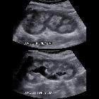
Toddler with
hypercalcemia. Sagittal US of the left and right kidneys shows echogenic medullary pyramids bilaterally and mild right hydronephrosis.The diagnosis was nephrocalcinosis and a mild left ureteropelvic junction obstruction.

Symptomatic
renal papillary varicosities and medullary nephrocalcinosis. Coronal CT image demonstrating patient’s focal nephrocalcinosis prior to treatment

Medullary
nephrocalcinosis • Medullary nephrocalcinosis - Ganzer Fall bei Radiopaedia

Preschooler
with chronic kidney disease. Sagittal US of the kidneys shows diffusely increased echogenicity throughout the medullary pyramids bilaterally.The diagnosis was nephrocalcinosis.


Rx de árbol
urinario que muestra múltiples calcificaciones milimétricas en topografía de las siluetas renales.

Familial
mixed nephrocalcinosis as a cause of chronic kidney failure: two case reports. Calcifications visible in the abdomen of patient 1. Images reveal mixed nephrocalcinosis with accentuated calcifications in the renal cortex and medulla. (A) X-ray. (B) Computed tomography (longitudinal cut). (C) Computed tomography (coronal cut).

Medullary
nephrocalcinosis • Medullary sponge kidney - Ganzer Fall bei Radiopaedia

Nephrocalcinosis
• Nephrocalcinosis - Ganzer Fall bei Radiopaedia

Acoustic
shadowing • Medullary nephrocalcinosis and renal calculi - Ganzer Fall bei Radiopaedia

Nephrocalcinosis
• Nephrocalcinosis - Ganzer Fall bei Radiopaedia

Nephrocalcinosis
• Nephrocalcinosis - Ganzer Fall bei Radiopaedia
Nephrocalcinosis, previous known as Anderson-Carr kidney or Albright calcinosis, refers to the deposition of calcium salts in the parenchyma of the kidney. It is divided into several types, with differing etiologies, based on the distribution:
- medullary nephrocalcinosis: 95%
- cortical nephrocalcinosis: 5%
- partial, combined cortical and medullary nephrocalcinosis : rare
History and etymology
An American surgeon L F Anderson in the 1940s, and a British radiologist, Reginald J Carr, in the 1950s, carried out pioneering work on the formation of renal calculi .
Siehe auch:
und weiter:


 Assoziationen und Differentialdiagnosen zu