Mesenchymales Hamartom

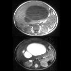
Toddler with
hepatomegalyAxial T1 weighted without contrast (above) and T2 weighted (below) MRI of the abdomen shows a mass in the right lobe as well as in the medial segment of the left lobe of the liver, measuring 13 x 13 x 7 cm in size and having a cystic and solid appearance.The diagnosis was mesenchymal hamartoma.

Hepatic
mesenchymal hamartoma • Hepatic mesenchymal hamartoma - Ganzer Fall bei Radiopaedia

Hepatic
mesenchymal hamartoma • Hepatic mesenchymal hamartoma - Ganzer Fall bei Radiopaedia

Hepatic
mesenchymal hamartoma • Hepatic mesenchymal hamartoma - Ganzer Fall bei Radiopaedia

Hepatic
mesenchymal hamartoma • Hepatic mesenchymal hamartoma - Ganzer Fall bei Radiopaedia

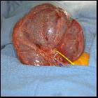
Toddler with
an abdominal mass. Surgical image of the under surface of the liver with the hila being dissected shows a large tumor in the left medial segment of the liver.The diagnosis was mesenchymal hamartoma.

Chondromesenchymal
hamartomas in a 24-year-old male mimicking a posterior mediastinal tumor and a 5-month-old boy with postoperative disseminated intravascular coagulation: two case reports. Radiological appearance of chondromesenchymal hamartoma of the chest wall in a 24-year-old adult. a, b DR suggested a left posterosuperior mediastinal mass and bronchitic changes. c CT revealed a benign expansile lesion in the posterior part of the left fifth rib with interior punctate calcifications. d-f MRI revealed a well-circumscribed expansile heterogeneous soft mass closely adjoining the fifth vertebral body in the left posterior mediastinum. DR, digital radiography; CT, computed tomography; DR, digital radiography; MRI, magnetic resonance imaging

Chondromesenchymal
hamartomas in a 24-year-old male mimicking a posterior mediastinal tumor and a 5-month-old boy with postoperative disseminated intravascular coagulation: two case reports. Imaging findings of chondromesenchymal hamartoma of the chest wall in a 5-month-old infant. a DR revealed a soft tissue mass in the left middle lung field, accompanied by collapse of the adjacent thoracic cage. b DR revealed postoperative loss of parts of the left fifth and sixth ribs. c, d CT revealed a benign solid-cystic lesion with multiple speckled and cord-like high-density shadows, arising from the axillary segment of the left fifth and sixth ribs; the corresponding cortical and medullary cavities of the ribs were involved. DR, digital radiography; CT, computed tomography
Mesenchymales Hamartom
Hamartom Radiopaedia • CC-by-nc-sa 3.0 • de
A hamartoma is a benign tumor-like malformation that consists of a collection of architecturally disorganized cells located in an area of the body where the cells are normally found. It is often due to abnormal development.
In radiology, hamartomas often mimic malignancy. Several hamartomata have characteristic imaging findings:
- biliary hamartomata
- breast hamartoma
- cardiac fibrous hamartoma
- fibrolipomatous hamartoma of the nerve
- gastrointestinal hamartomatous polyps
- hepatic mesenchymal hamartoma
- hypothalamic hamartoma
- pulmonary hamartoma
- splenic hamartoma
- subependymal hamartoma
History and Etymology
The term hamartoma derives from the Greek word hamartía meaning error or failure.
Related Radiopaedia articles
Curricula
- curriculum
- anatomy curriculum
- imaging curricula
- breast curriculum
- cardiac curriculum
- central nervous system curriculum
- chest curriculum
- gastrointestinal curriculum
- gynecology curriculum
- head and neck curriculum
- hepatobiliary curriculum
- intervention curriculum
- musculoskeletal curriculum
- obstetric curriculum
- pediatric curriculum
- post-mortem and forensic curriculum
- urogenital curriculum
- vascular curriculum
- medical student curriculum
- pathology curriculum
- general pathology
- systemic pathology
- pathology of the vascular system
- cardiac pathology
- pathology of the hematological and lymphatic systems
- pathology of the lung and pleura
- pathology of the head and neck
- pathology of the gastrointestinal system
- pathology of the liver, biliary tract and pancreas
- pathology of the kidney and urinary tract
- pathology of the male genital system
- pathology of the female genital system
- pathology of the breast
- pathology of the endocrine system
- pathology of the skin
- pathology of the musculoskeletal system and soft tissues
- pathology of the nervous system
- pathology of the ocular system
- physics curriculum
- radiography curriculum
- general radiography curriculum
- CT curriculum
- MRI
- ultrasound
- nuclear medicine
- radiation therapy
Siehe auch:

 Assoziationen und Differentialdiagnosen zu Mesenchymales Hamartom:

