Pfeiffer syndrome

Prenatal
diagnosis of pfeiffer syndrome type II using ultralow dose CT. Magnetic resonance imaging (half-Fourier acquisition single-shot turbo spin-echo) of the fetal head at 31 3/7 weeks of gestation. Coronal image shows a cloverleaf skull with temporal protrusion (arrows) and mild ventriculomegaly (asterisk).

Prenatal
diagnosis of pfeiffer syndrome type II using ultralow dose CT. Sagittal image shows proptosis (arrowhead) and mild ventriculomegaly (asterisk).

Prenatal
diagnosis of pfeiffer syndrome type II using ultralow dose CT. Three-dimensional computed tomography of the fetus at 32 3/7 weeks of gestation. Volume rendering of full skeletal image shows craniosynostosis, the large distal phalanx (arrowhead) of the left great toe, and bilateral radio-ulnar-humeral synostoses (arrows).

Prenatal
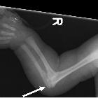
diagnosis of pfeiffer syndrome type II using ultralow dose CT. Maximum intensity projection of the left upper extremity shows radio-ulnar-humeral synostosis (arrow).

Prenatal
diagnosis of pfeiffer syndrome type II using ultralow dose CT. Maximum intensity projection image of the right foot also shows large distal phalanges (arrowhead) of the great toe and triangular hypoplastic first proximal phalanx (arrow), as well as volume rendering.

Prenatal
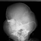
diagnosis of pfeiffer syndrome type II using ultralow dose CT. Anterior−posterior view of the head shows a cloverleaf skull.

Prenatal
diagnosis of pfeiffer syndrome type II using ultralow dose CT. Plain radiographs of the right upper extremity shows radio-ulnar-humeral synostosis (arrow).

Prenatal
diagnosis of pfeiffer syndrome type II using ultralow dose CT. Plain radiograph of the feet shows broad great toes (arrowheads) and triangular hypoplastic first proximal phalanges (arrows) as well as fetal computed tomography images.

Kleeblattschädel
skull presenting in concert with Pfeiffer syndrome. a Patient with tri-lobar skull. b A side angle of the tri-lobar skull

Kleeblattschädel
skull presenting in concert with Pfeiffer syndrome. CAT showing gross biventricular hydrocephalus with disproportionate ballooning of the temporal horns

Kleeblattschädel
skull presenting in concert with Pfeiffer syndrome. 3D reconstruction imaging of the skull

Cloverleaf
skull (craniosynostosis) • Pfeiffer syndrome - Ganzer Fall bei Radiopaedia

Pfeiffer
syndrome • Pfeiffer syndrome - craniosynostosis - Ganzer Fall bei Radiopaedia
Pfeiffer syndrome (also known as type V acrocephalosyndactyly) is characterized by skull and limb abnormalities.
Epidemiology
It affects about 1 in 100,000 births
Clinical presentation
- craniosynostosis
- hypertelorism
- proptosis
- maxillary hypoplasia
- brachydactyly
- syndactyly
Pathology
Pfeiffer syndrome is strongly associated with mutations of the fibroblast growth factor receptor 1 gene (FGFR1) on chromosome 8 or the fibroblast growth factor receptor 2 (FGFR2) gene on chromosome 10.
Classification
- type 1: classic Pfeiffer; most individuals have normal intelligence and lifespan. Inherited in an autosomal dominant pattern.
- type 2: includes a cloverleaf skull (Kleeblattschädel); occurs sporadically and has a poor prognosis with severe neurological compromise.
- type 3: includes craniosynostosis and severe proptosis; occurs sporadically and has poor prognosis.
History and etymology
It was first described by Rudolph Arthur Pfeiffer in 1964.
Differential diagnosis
Consider other forms of acrocephalosyndactyly such as
Siehe auch:
und weiter:

 Assoziationen und Differentialdiagnosen zu Pfeiffer-Syndrom:


