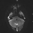
Kleinhirnabszess

Dual rim sign
(brain abscess) • Dual rim sign (annotated image) - Ganzer Fall bei Radiopaedia

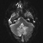
Cerebellar
abscess secondary to occipital dermoid cyst with dermal sinus. Sagittal View MRI Brain, T2, FLAIR images showing perilesional oedema in cerebellum and communication.

Cerebellar
abscess secondary to occipital dermoid cyst with dermal sinus. Pre contrast T1W image shows multiple foci in posterior and midline region of cerebellar folia in keeping with leakage of fat content from high signal dermoid cyst in occipital region of scalp through sinus.

Cerebellar
abscess secondary to occipital dermoid cyst with dermal sinus. Pre contrast T1W image shows multiple foci in posterior and midline region of cerebellar folia in keeping with leakage of fat content from high signal dermoid cyst in occipital region of scalp through sinus.

Cerebellar
abscess secondary to occipital dermoid cyst with dermal sinus. Pre-contrast T1W coronal images – high signal in subarachnoid space in posterior fossa consistent with leakage of fatty content of dermoid.
Cerebellar
abscess secondary to occipital dermoid cyst with dermal sinus. MRI Brain, axial view, diffusion weighted image showed restricted diffusion.
Cerebellar
abscess secondary to occipital dermoid cyst with dermal sinus. MRI Brain, axial view, diffusion weighted image showed restricted diffusion.

Cerebellar
abscess secondary to occipital dermoid cyst with dermal sinus. Axial T2W image, lesion in midline with perilesional oedema and mass effect. Communication/dermal sinus tract through occipital bone defect to scalp dermoid with secondary infection. Incidental detected right sided acoustic schwannoma.

Brain abscess
• Cerebral abscesses - Ganzer Fall bei Radiopaedia

Cerebellar
abscess secondary to occipital dermoid cyst with dermal sinus. Post-contrast coronal T1W images showed ring enhancement around focal collection.

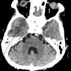
Cerebral
abscess • Cerebral abscess - Ganzer Fall bei Radiopaedia

Cerebral
venous thrombosis, subdural empyema and cerebral abscess as complications of coalescent otomastoiditis. Axial CT shows small hypodense area in the right temporal lobe, which should prompt the suspicion of cerebral abscess in the given context.

Cerebral
venous thrombosis, subdural empyema and cerebral abscess as complications of coalescent otomastoiditis. MRI T2 shows small delineated hyperintense lesion with surrounding diffuse peripheral hyperintensity in the right temporal lobe, compatible with cerebral abscess with surrounding oedema.

Cerebral
venous thrombosis, subdural empyema and cerebral abscess as complications of coalescent otomastoiditis. B1000 and ADC images with small area of restricted diffusion in right temporal lobe.

Cerebral
venous thrombosis, subdural empyema and cerebral abscess as complications of coalescent otomastoiditis. MRI 3D reconstruction shows small peripherally enhancing area in the right temporal lobe, compatible with cerebral abscess.

Dual rim sign
(brain abscess) • Brain abscess with dual rim sign - Ganzer Fall bei Radiopaedia

Dual rim sign
(brain abscess) • Dual rim sign (brain abscess) - Ganzer Fall bei Radiopaedia

Cerebellar
abscess secondary to occipital dermoid cyst with dermal sinus. Post-contrast T1W MR shows high signal foci which were non-enhancing suggesting leakage of fat containing dermoid cyst in cerebellar folia.

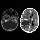
Hirnabszess
in der MRT (axial T1w nach Kontrastmittel) mit klassischer ringförmiger Anreicherung und deutlichem perifokalem Ödem.
School ager
with pus draining from the right ear. Axial CT with contrast of the brain with bone windows (left) shows opacification and destruction of the right mastoid air cells while axial CT with contrast of the brain with soft tissue windows (right) shows a large low density ring enhancing lesion in the right cerebral hemisphere that is causing midline shift to the left.The diagnosis was right coalescent mastoiditis with an intracranial abscess.

Brain abscess
• Cerebral abscess with ventriculitis - Ganzer Fall bei Radiopaedia

Brain abscess
• Brain abscess with ventriculitis - Ganzer Fall bei Radiopaedia

Brain abscess
• Brain abscess - Ganzer Fall bei Radiopaedia

Brain abscess
• Skull base osteomyelitis and temporal lobe abscess - Ganzer Fall bei Radiopaedia

Brain abscess
• Fungal cerebral abscesses - Ganzer Fall bei Radiopaedia

Brain abscess
• Cerebellar abscess - Ganzer Fall bei Radiopaedia

Cerebral ring
enhancing lesions • Cerebral abscess - Ganzer Fall bei Radiopaedia

Brain abscess
• Cerebral abscess and ventriculitis - Ganzer Fall bei Radiopaedia

Endopthalmitis,
brain abscesses probably seconday to metastatic renal abscess secondary to virulent Klebsiella species. CT head with contrast shows multiple rim enhancing parenchymal lesions with central low attenuation. Findings consistent with cerebral abscesses.

Brain abscess
• Cerebral abscess - Ganzer Fall bei Radiopaedia

Brain abscess
• Cerebral abscess - Ganzer Fall bei Radiopaedia

Brain abscess
• Cerebral abscess - Ganzer Fall bei Radiopaedia

Brain abscess
• Cerebral abscesses - Ganzer Fall bei Radiopaedia
Endopthalmitis,
brain abscesses probably seconday to metastatic renal abscess secondary to virulent Klebsiella species. CT head with contrast shows multiple rim enhancing parenchymal lesions with central low attenuation. Findings consistent with cerebral abscesses.

Cerebellar
abscess secondary to occipital dermoid cyst with dermal sinus. Sagittal T2, FLAIR images showing perilesional oedema in cerebellum and communication.

Endopthalmitis,
brain abscesses probably seconday to metastatic renal abscess secondary to virulent Klebsiella species. CT head with contrast shows multiple rim enhancing parenchymal lesions with central low attenuation. Findings consistent with cerebral abscesses.

Cerebellar
abscess secondary to occipital dermoid cyst with dermal sinus. Post-contrast coronal T1W images showed ring enhancement around focal collection.
Kleinhirnabszess
Siehe auch:
- Hirnmetastase
- Glioblastoma multiforme
- Hirnabszess
- Sinusthrombose
- Metastase
- Demyelinisierende Erkrankung
- intrakranielle Echinokokkose
- Läsionen mit ringförmiger Kontrastmittelanreicherung
- cerebellar tuberculoma
- septisch-embolische Herdenzephalitis
- Strahlennekrose
- haemorrhage
- dermal sinus tract
- septisch-metastatische Herdenzephalitis
und weiter:


 Assoziationen und Differentialdiagnosen zu Kleinhirnabszess:


septisch-embolische
Herdenzephalitis
