growing skull fracture


Leptomeningeal cysts, also known as growing skull fractures, are an enlarging skull fracture that occurs near post-traumatic encephalomalacia. The term cyst is actually a misnomer, as it is not a cyst, but an extension of the encephalomalacia. Hence, it is usually seen a few months post-trauma.
Epidemiology
The majority occur in children <3 years. They complicate ~1% of skull fractures .
Clinical presentation
Children can present with :
Pathology
The exact pathogenesis remains unclear but it is thought they occur secondary to skull fractures causing dural tears allowing the leptomeninges and/or cerebral parenchyma to herniate into it . Pulsations from CSF erode the fracture margin, resulting in eventual expansion and non-union .
Radiographic features
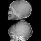
Plain radiograph
- round or oval lucency with smooth margins
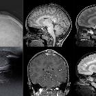
CT
CT scan is the modality of choice for the evaluation of leptomeningeal cyst. It consists of a lytic calvarial lesion with scalloped edges, in which encephalomalacia invaginates. The following features may also be present :
- extracranial brain herniation
- hydrocephalus
- unilateral ventricular dilatation
- porencephalic cyst
Differential diagnosis
- eosinophilic granuloma
- calvarial metastases
- epidermoid cyst
- osteomyelitis
- congenital calvarial defect
Siehe auch:
- Schädelfraktur
- Knochenmetastasen des Schädels
- intraossäre Epidermoidzyste
- eosinophiles Granulom des Schädels
- Osteomyelitis des Schädels
- kongenitaler Kalottendefekt
und weiter:


 Assoziationen und Differentialdiagnosen zu wachsende Schädelfraktur: