dural arteriovenous shunts

Location,
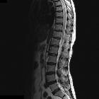
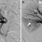
length, and enhancement: systematic approach to differentiating intramedullary spinal cord lesions. Dural arteriovenous fistula. A 50-year-old male with gradual onset of inability to walk, off and on constipation, and urinary incontinence. a Axial T2 image demonstrates hyperintensity in the cord and multiple serpiginous flow voids (arrow) within the thecal sac. b Sagittal T2 image reveals long segment involvement (bracket). c Conventional spinal angiogram shows injection of a paraspinal artery with simultaneous robust filling of a corkscrew venous vessel

Dural
falco-tentorial arteriovenous fistula. Volume rendering image depicts dilated extracranial arteries that are branches originating from the external carotid artery and supplying the lesion.

Dural
falco-tentorial arteriovenous fistula. T1 VIBE after gadolinium enhancement axial image shows hypointense area due to Onyx and reveals little and persistent anterior arterial supply.

Dural
falco-tentorial arteriovenous fistula. Lateral right internal carotid angiogram in venous phase confirms the vein flux anomaly.

Dural
falco-tentorial arteriovenous fistula. Frontal right internal carotid angiogram in venous phase reveals absence of internal venous flow due to its early drain to cavernous sinus via lenticulostriate vein.

Dural
falco-tentorial arteriovenous fistula. Lateral fluoroscopy view of Onyx treatment.

Dural
falco-tentorial arteriovenous fistula. Lateral left internal carotid angiogram confirms posterior meningeal artery supply.

Dural
falco-tentorial arteriovenous fistula. Frontal fluoroscopy view of Onyx embolic material within arterio-venous dural fistula.

Dural
falco-tentorial arteriovenous fistula. Frontal left internal carotid angiogram depicts posterior meningeal artery supply with anomalous ascending pharyngeal artery origin in internal carotid. Early vein drain and vein reflux are also demonstrated.

Dural
falco-tentorial arteriovenous fistula. Lateral left occipital artery angiogram depicts a falcine arterial supply to the fistula and early venous drain.

Dural
falco-tentorial arteriovenous fistula. Frontal left occipital carotid angiogram shows transforaminal supply to the fistula.

Dural
falco-tentorial arteriovenous fistula. Lateral right external carotid angiogram supports the middle meningeal artery supply with drainage to Gallen vein and straight sinus with internal cerebral veins reflux.

Dural
falco-tentorial arteriovenous fistula. Frontal right external carotid angiogram demostrates supplies from the middle meningeal artery.

Dural
falco-tentorial arteriovenous fistula. T2 blade axial image with hypointense tubular structures corresponding with vascular malformation.

Dural
falco-tentorial arteriovenous fistula. T2 blade axial image with hypointense tubular structures corresponding with vascular malformation.

Dural
falco-tentorial arteriovenous fistula. Major Intensity Projection (MIP) image shows clustered vessels surrounding straight sinus and Gallen vein, accompanied by venous ectasia.

Dural
falco-tentorial arteriovenous fistula. Axial CT angiography demonstrates the presence of a vascular malformation.

Location,
length, and enhancement: systematic approach to differentiating intramedullary spinal cord lesions. Dural arteriovenous fistula. A 72-year-old male with progressive lower extremity weakness, without improvement in symptoms after lumbar laminectomy. a Axial T2 image demonstrates numerous flow voids in the thecal sac (arrowhead). There is high signal in the cord due to congestion (arrow). b There is long-segment high T2 signal in the cord, with serpiginous vessels in the posterior thecal sac (bracket). c Spinal angiogram shows the right T4–T5 supreme intercostal artery as a feeding vessel. d MRA confirms the arteriovenous connection

Dural
falco-tentorial arteriovenous fistula. Non-contrast CT axial shows enlargement of vascular foramina in the skull.

Dural
falco-tentorial arteriovenous fistula. Non-contrast CT axial image discloses an spontaneous hyperdense tubular mass in the falx causing incipient hydrocephalus.

Dural
arteriovenous fistula • Dural arteriovenous fistula - Ganzer Fall bei Radiopaedia

Dural
arteriovenous fistula • Intracranial dural arteriovenous fistula - Ganzer Fall bei Radiopaedia

Dural
arteriovenous fistula • Dural arteriovenous fistula (dAVF) - progression from normal to marked - Ganzer Fall bei Radiopaedia

Dural
arteriovenous fistula • Dural arteriovenous fistula - Ganzer Fall bei Radiopaedia

Dural
arteriovenous fistula • Posterior cranial fossa dural arteriovenous fistula - pulsatile tinnitus - Ganzer Fall bei Radiopaedia

Dural
arteriovenous fistula • Dural arteriovenous fistula - Ganzer Fall bei Radiopaedia

Dural
arteriovenous fistula • Anterior cranial fossa dural arteriovenous fistula - Ganzer Fall bei Radiopaedia

Dural
arteriovenous fistula • Intracranial dural arteriovenous fistula - Ganzer Fall bei Radiopaedia

Dural
arteriovenous fistula • Dural arteriovenous fistula - Ganzer Fall bei Radiopaedia

Dural
arteriovenous fistula • Indirect caroticocavernous fistula - Ganzer Fall bei Radiopaedia

Dural
arteriovenous fistula • Dural arteriovenous fistula - posterior fossa - Ganzer Fall bei Radiopaedia

Dural
arteriovenous fistula • Dural arteriovenous fistula - anterior cranial fossa - Ganzer Fall bei Radiopaedia

Dural
falco-tentorial arteriovenous fistula. SWI axial image depicts hypointense area secondary to Onyx.
Dural arteriovenous shunts (DAVS) are rare congenital arteriovenous malformations (CAVMs). On the basis of clinical and anatomical features DAVS have three different types:
- dural sinus malformations (DSMs)
- infantile or juvenile DAVS (IDAVS)
- adult DAVS (ADAVS)
Siehe auch:
- Arteria carotis interna
- Sinus sigmoideus
- Arteriovenöse Malformation
- Subarachnoidalblutung
- subdurales Hämatom
- Sinus transversus
- Sinus sagittalis superior
- Sinus cavernosus
- Sinus rectus
- Cognard classification of dural arteriovenous fistulas
- spinale durale arteriovenöse Fistel
- intrakranielle vaskuläre Malformationen
- Borden classification of dural arteriovenous fistulas
- Carotis-Sinus-cavernosus-Fistel
- Arteriovenöse Fistel
und weiter:
- zerebrale arteriovenöse Malformation
- Intrakranielle Blutung
- neuroradiologisches Curriculum
- intramedulläre spinale Tumoren
- Varizen der Orbita
- pulssynchrones Ohrgeräusch
- spinal AVM classification
- cerebral malformation
- Blue-rubber-bleb-naevus-Syndrom
- Onyx
- Klassifikation zerebraler vaskulärer Malformationen
- kraniale durale AV-Fistel
- dural arteriovenous fistula: posterior fossa
- durale AV Fistel Einteilung
- Malformationen der duralen Sinus
- spontanes subdurales Hämatom

 Assoziationen und Differentialdiagnosen zu durale AV-Fistel:
intrakranielle
vaskuläre Malformationen


