jejunale Diverticulose

Dünndarmdivertikulitis
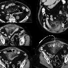
mit Perforation und Abszess. Links oben zahlreiche Divertikel des Dünndarms, an dieser Stelle reizlos. Links Mitte deutliche Wandverdickung weiter kaudal gelegener Dünndarmabschnitte. Links unten große Abszessformation (ca. 7 cm) zwischen den Dünndarmschlingen auf Beckeneingangsebene ventral. Rechts oben koronare Darstellung von Dünndarmdivertikel links im Bauch ohne Perforation, im Mittelbauch Abszessformation mit deutlich entzündlich veränderten angrenzenden Dünndarmschlingen. Rechts unten Z.n. Drainage der Abszessformation, bei der ca. 50 ml Pus abgezogen werden konnten.

Zahlreiche
Dünndarmdivertikel, z.T. sehr groß, im wesentlichen im Jejunum, wiederholte Perforation


Jejunal
diverticulosis. 1,5 meter of distended jejunum with multiple diverticula was resected

Massive lower
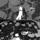
gastrointestinal bleeding from jejunal diverticulum with an occult Dieulafoy’s lesion. Coronal (upper) and axial (lower) images. Swirling jet of contrast material (black arrow) within the lumen of the diverticulum (white arrow). A collapsed inferior cava vein indicates hypovolaemia (white arrowhead).

Massive lower
gastrointestinal bleeding from jejunal diverticulum with an occult Dieulafoy’s lesion. Coronal (upper) and axial (lower) images. The amount of intraluminal contrast increases in diverticular (black arrowhead) and jejunal (black arrow) lumen in comparision with arterial phase. Hyperattenuating material in the colon suggests blood (white arrowhead).

Jejunoileal
diverticulitis • Jejunal diverticulitis - acute - Ganzer Fall bei Radiopaedia

Jejunoileal
diverticula • Jejunal diverticulosis - Ganzer Fall bei Radiopaedia

Jejunal
diverticulosis. Axial image at level Th12. Pneumatosis intestinalis in the diverticular wall (arrows).

Jejunoileal
diverticulitis • Jejunal diverticulitis - Ganzer Fall bei Radiopaedia

Jejunoileal
diverticula • Jejunoileal diverticulosis - Ganzer Fall bei Radiopaedia

Jejunoileal
diverticula • Jejunal diverticulitis with migration of a large, obstructing enterolith - Ganzer Fall bei Radiopaedia

Jejunoileal
diverticulitis • Terminal ileal diverticulitis - Ganzer Fall bei Radiopaedia

Jejunoileal
diverticulitis • Jejunal diverticulitis - Ganzer Fall bei Radiopaedia

Jejunoileal
diverticula • Jejunoileal diverticula - Ganzer Fall bei Radiopaedia

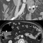
Massive lower
gastrointestinal bleeding from jejunal diverticulum with an occult Dieulafoy’s lesion. Coronal (upper) and axial (lower) images. Diverticulum (white arrow) originating in the jejunum wall (*), with stranding of the adjacent fat (white arrowhead). There was no hyperattenuating material within the diverticulum lumen.

Duodenal
diverticulum • Small bowel diverticulosis - Ganzer Fall bei Radiopaedia
Jejunoileal diverticula, also referred to as jejunal diverticula or diverticulosis as most of the diverticula are located in the jejunum, are outpouchings from the jejunal and ileal wall on their mesenteric border that represent mucosal herniation through sites of wall weakening .
Jejunoileal diverticulitis is much rarer than colonic diverticulitis.
See also
Please refer to the articles on duodenal diverticula and on Meckel diverticulum for a discussion of other small intestine diverticular disease.
Siehe auch:
- Duodenaldivertikel
- Meckel-Divertikel
- Divertikulose
- juxtapapilläres Duodenaldivertikel
- Dünndarmdivertikulitis
- Perforation eines Duodenaldivertikels
- blutendes Jejunaldivertikel
- Divertikel des Gastrointestinaltraktes
und weiter:

 Assoziationen und Differentialdiagnosen zu Dünndarmdivertikel:


