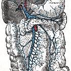
gallbladder

The gallbladder is a pear-shaped musculomembranous sac located along the undersurface of the liver. It functions to accumulate and concentrate bile between meals.
Gross anatomy
Macroscopic
The normal adult gallbladder measures from 7-10 cm in length and 3-4 cm in transverse diameter . The gallbladder communicates with the rest of the biliary system by way of the cystic duct, with bidirectional drainage of bile to and from the common hepatic duct.
For descriptive purposes, it may be divided into the following segments :
- fundus
- body
- infundibulum: tapered segment between body and neck
- Hartmann pouch: small outpouching, variably identified, at the infundibulum
- neck: communicates with the cystic duct
The gallbladder is closely apposed to the liver within the fossa. Indeed, the liver's serosal covering (visceral peritoneum) extends over and completely covers the free surface of the gallbladder . The gallbladder connects to the liver via a layer of dense connective tissue (adventitia), which contains small draining cystic veins, autonomic innervation, lymphatic drainage, and variable accessory bile ducts (of Lushka) . In some cases, the gallbladder "hangs" from the liver from a short mesentery of redundant connective tissue .
Microscopic
The gallbladder wall comprises :
- serosa (visceral peritoneum): only covering the inferior free surfaces of the gallbladder
- muscular outer layer (muscularis propria or externa)
- lamina propria
- mucosa (single cell layer)
Unlike other foregut-derived organs, the lamina propria and muscular layers are directly apposed because there are no submucosal or muscularis mucosae layers .
The outer muscular layer forms the framework of the gallbladder and consists of dense fibrous tissue interlaced with randomly-oriented smooth muscle fibers, contrasting with the well-organized longitudinal and circular organization within the intestine .
The inner mucosal layer consists of branching folds of lamina propria covered in a single-cell layer of columnar epithelium, overall lending the appearance of minute rugae . There are extensive capillaries and small venules, but absent lymphatics . Mucus-secreting glands are only present at in the lamina propria layer at the gallbladder neck, and may be joined by enteroendocrine cells .
Rokitansky-Aschoff sinuses are deep outpouchings or diverticula of the mucosal layer that extend into the outer muscular layer and are variably present . They are the structure implicated in adenomyomatosis, and are noted in more than half of cases of chronic cholecystitis .
Location
The gallbladder is located in a shallow fossa along the inferior aspect of the liver, in line with the interlobar fissure that separates right and left liver lobes. It has an oblique craniocaudal/anterolateral lie, such that the neck is located to the right of the porta hepatis and the fundus directed inferiorly to the anterior border of the right liver lobe. The fundus commonly projects inferior to the right liver margin.
Most frequent aberrant locations in descending order are beneath left lobe of liver, intraheptic, retrohepatic, within the falciform ligament, within the interlobar fissure, suprahepatic, and within the anterior abdominal wall.
Relations
- superiorly: visceral surface of the liver, anterior abdominal wall
- inferiorly: transverse colon, second part of the duodenum (or pylorus of the stomach)
- anteriorly: visceral surface of the liver, transverse colon, 9 costal cartilage
- posteriorly: right kidney, distal first part and proximal second part of the duodenum
- medially: first part of the duodenum, free margin of the lesser omentum and epiploic foramen
- laterally: right lobe of the liver
Function
The gallbladder is involved in the storage, concentration, and ejection of the bile.
The adult gallbladder holds ~30-50 mL of bile when distended , although if obstructed can distend to accommodate up to 300 mL .
The gallbladder concentrates bile using mechanism of active transport of sodium and chloride, effectively removing water and slightly increasing acidity of bile. The net effect is a 10-fold increase in bile salt concentration during storage .
In response to the detection of ingested fat, gallbladder contraction is signaled by way of a neurohormonal pathway that results in prompt excretion of the biliary payload.
Arterial supply
The gallbladder receives the vast majority of its arterial blood from the cystic artery.
Venous drainage
There is no single cystic vein, but rather the gallbladder drains directly into the venous system of the liver through the gallbladder fossa (cystic veins) and by a number of veins into the right branch of the portal vein .
Lymphatic drainage
Gallbladder lymphatic drainage is complex. Three distinct pathways have been described based on cadaveric dissection :
- cholecysto-retropancreatic: following common duct inferiorly to a retroportal node posterior to pancreatic head (primary pathway)
- cholecysto-celiac: via hepatoduodenal ligament to celiac nodes
- cholecysto-mesenteric: anterior to portal vein to superior mesenteric root nodes
These are thought to converge at aortocaval and para-aortic nodes near the renal veins .
Innervation
The gallbladder receives both sympathetic and vagal supply:
- sympathetic: via the celiac plexus
- vagal: via the hepatic branches of anterior vagal trunk
Variant anatomy
The gallbladder has a number of variations in its anatomy based on:
- morphology
- fold (common)
- Phrygian cap: fundus folded back upon itself - primary significance is to avoid confusion
- sigmoid gallbladder
- septation (uncommon)
- congenital or acquired (secondary to chronic cholecystitis)
- single or multiple
- any orientation
- Hartmann pouch (infundibulum)
- in some instances, the neck is focally dilated (adjacent to the body)
- probably pathological, related to cholelithiasis
- floating gallbladder
- gallbladder may possess a peritoneal mesentery
- may predispose to gallbladder torsion
- diverticula (rare)
- containing all layers of the gallbladder wall (vs Rokitansky-Aschoff sinuses)
- fold (common)
- number
- location: ectopic gallbladder has been reported in many different abdominal sites and can result in increased complexity when undertaking cholecystectomy
- intrahepatic
- retrohepatic
- transverse
- retroperitoneal
- left-sided: extremely rare (<0.2%)
- located to the left of the falciform ligament without situs inversus
- normally not diagnosed on preoperative imaging (i.e. apparent only at operation)
Related pathology
- cholelithiasis
- cholecystitis and related conditions
- gallbladder polyps
- gallbladder carcinoma
- adenomyomatosis
- microgallbladder
Siehe auch:
- Phrygische Mütze der Gallenblase
- Gallenblasenkarzinom
- Adenomyomatose
- Vena portae
- akute Cholezystitis
- emphysematöse Cholezystitis
- Porzellangallenblase
- Polypen der Gallenblase
- Cholezystolithiasis
- strawberry gallbladder
- Mirizzi-Syndrom
- Gallenblasenduplikatur
- Pseudodivertikel der Gallenblase
- Gallenblasenfunktionstest
- Leberpforte
und weiter:

 Assoziationen und Differentialdiagnosen zu Gallenblase:


