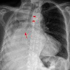
vanishing lung syndrome


Idiopathic giant bullous emphysema, also known as vanishing lung syndrome (VLS), is characterized by giant emphysematous bullae, which commonly develop in the upper lobes and occupy at least one-third of a hemithorax. It is a progressive condition that is also associated with several forms of emphysema.
Epidemiology
Idiopathic giant bullous emphysema is a rare syndrome that differs from conventional forms of bullous emphysema; affecting a younger population, most commonly young men.
Associations
Most affected patients smoke cigarettes.
The condition has been associated with:
Clinical presentation
Affected patients may be asymptomatic or experience hypoxia, severe dyspnea and/or chest pain.
Pathology
Location/distribution
The bullae commonly involve the upper lobes with an asymmetric distribution and paraseptal location.
Radiographic features
Plain radiograph
Plain radiographic features can be non-specific:
- bullae occupy more than one-third of the affected hemithorax (vary in size 1-20 cm)
- upper lobes have greater involvement
- there is bilateral and asymmetric lung involvement
- may have mass effect on adjacent structures (lung parenchyma atelectasis, invert the ipsilateral diaphragm or contralateral displacement of the mediastinum and displacement of junction lines)
CT
High-resolution chest CT is used to assess the extent of disease and determine
suitability for lung volume reduction surgery. Features on CT include:
- bullae predominantly in the subpleural location
- the size of bullae ranges from 1 to 20 cm in diameter, but most measure 2-8 cm
- usually asymmetric, with one lung involved to a greater extent than the other
- the presence of concomitant foci of paraseptal and centrilobular emphysema
Treatment and prognosis
The condition tends to be progressive. Criteria for bullectomy include large bullae with significant symptoms (reduced lung function or infection).
Complications
- compression of surrounding lung (compressive atelectasis)
- pneumothorax
- bullae infection
- higher risk of lung cancer
History and etymology
In 1937, Burke described a case of “vanishing lungs” in a 35-year-old man who had progressive dyspnea, respiratory failure, and radiographic and pathologic findings of giant bullae that occupied two-thirds of both hemithoraces.
Siehe auch:
- Pneumothorax
- Lungenkarzinom
- Lungenemphysem
- einseitig vermehrte Transparenz Thorax
- Ehlers-Danlos syndrome
- Marfan-Syndrom
- Alpha-1-Antitrypsin-Mangel
- paraseptales Emphysem
- bullöses Emphysem
- Emphysem
- pulmonale Bullae
und weiter:

 Assoziationen und Differentialdiagnosen zu idiopathisches Lungenemphysem mit riesigen Bullae:


