Epididymal mass

Adenomatoid
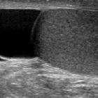
tumour of epididymis: ultrasonographic evaluation with surgical correlation. A split screen composite gray scale ultrasound showing a rounded solid mass of rather high echogenicity in continuity with the lower pole of the testis. A small cyst can be seen in the mass.

Epididymal
haematoma: multiparametric ultrasonographic findings.. Long axis grey-scale image showing the circumscribed hypoechoic lesion with some internal echogenic septations and debris which was situated in the right paratesticular tissues.

Epididymal
haematoma: multiparametric ultrasonographic findings.. Contrast-enhanced ultrasonographic image shows that there is no sign of vascularity within the lesion. There is mildly increased vascularity at the periphery of the lesion.

Epididymal
haematoma: multiparametric ultrasonographic findings.. Colour-coded elastographic image shows the lesion to be predominantly homogeneously soft except for the part where multiple colours can be seen. (Blue colour represents soft and red colour represents hard tissue).
Epididymal lesions are most commonly encountered on ultrasonography. Most epididymal lesions are benign; malignant lesions are rare.
They can comprise of
Benign solid lesions
- adenomatoid tumor of the scrotum: most common epididymal mass
- epididymal leiomyoma
- papillary cystadenoma of the epididymis
- sperm granulomas / post-vasectomy granuloma
- sarcoid - epidymal sarcoidosis
- epididymal tuberculosis
Benign cystic lesions
Malignant lesions
- epididymal metastases
- epididymal leukemia
- epididymal leiomyosarcoma
See also
Siehe auch:
- Epididymitis
- Spermatozele
- Nebenhodenzysten
- Zystadenom des Nebenhodens
- sperm granulomas
- post vasectomy granuloma
- epididymal lipoma
- adenomatoid tumours of the scrotum
- epididymal sarcoidosis
und weiter:

 Assoziationen und Differentialdiagnosen zu Tumoren des Nebenhodens:


